Showing posts with label P450. Show all posts

Saturday, May 24, 2014

[Fe3S4] ferredoxin from Rhodopseudomonas palustris

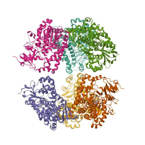
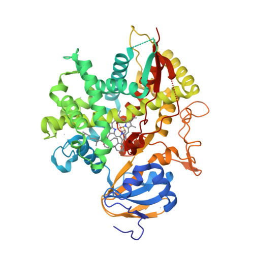
The crystal structure of a novel [Fe3S4] ferredoxin associated with CYP194A4 from Rhodopseudomonas palustris has been solved at 2.15 Å resolution [1—3]. The ferredoxin, HaPuxC, contains an atypical CXXHXXC(X)nCP iron-sulphur cluster-binding motif. HaPuxC is the first P450 electron-transfer partner of this type to be structurally characterised.

  1. Zhang, T., Zhang, A., Bell, S.G., Wong, L.-L. and Zhou, W. (2014) The structure of a novel electron-transfer ferredoxin from Rhodopseudomonas palustris HaA2 which contains a histidine residue in its iron-sulfur cluster-binding motif. Acta Crystallographica D70, 1453—1464.
  2. PDB:4ID8
  3. PDB:4OV1

Wednesday, May 15, 2013

Aldosterone synthase structures

Earlier this year [1], the crystal structures of human aldosterone synthase (CYP11B2) were solved in complex with a substrate 11-deoxycorticosterone [2] and an inhibitor fadrozole [3].

  1. Strushkevich, N., Gilep, A.A., Shen, L., Arrowsmith, C.H., Edwards, A.M., Usanov, S.A. and Park, H.-W. (2013) Structural insights into aldosterone synthase substrate specificity and targeted inhibition. Molecular Endocrinology 27, 315—324.
  2. PDB:4DVQ
  3. PDB:4FDH

Thursday, February 28, 2013

The first CYP1A1 structure

CYP1A1 was one of the first P450 enzymes to be characterised and, as its name indicates, holds the first place in the systematic nomenclature of P450s [1]. However, it was not until last year that the first crystal structure of human CYP1A1 in complex with α-naphthoflavone has been determined at 2.6 Å resolution [2]. The structure [3] is released this week.

  1. Nebert, D.W., Nelson, D.R., Coon, M.J., Estabrook, R.W., Feyereisen, R., Fujii-Kuriyama, Y., Gonzalez, F.J., Guengerich, F.P., Gunsalus, I.C., Johnson, E.F., Loper, J.C., Sato, R., Waterman, M.R. and Waxman, D.J. (1991) The P450 superfamily: update on new sequences, gene mapping, and recommended nomenclature. DNA Cell Biol. 10, 1—14.
  2. Walsh, A.A., Szklarz, G.D. and Scott, E.E. (2012) Cytochrome P450 1A1 structure and utility in predicting drug and xenobiotic metabolism. Proceedings of the 19th International Symposium on Microsomes and Drug Oxidations and 12th European ISSX Meeting, Noordwijk, The Netherlands, 17—21 June 2012.
  3. PDB:4I8V

Thursday, May 10, 2012

P450-flavodoxin fusion enzyme XplA

XplA is a P450-flavodoxin fusion enzyme that mediates the metabolism of the military explosive RDX (1,3,5-trinitro-1,3,5-triazinane) in Rhodococcus rhodochrous 11Y [1]. Bui et al. have conducted a detailed spectroscopic and crystallographic study of this unusual hemoflavoprotein [2, 3].

The XplA P450 has evolved as a reductase (rather than oxidase) of RDX and structural alterations to its heme- and FMN-binding domains have led to reduction potentials for low-spin heme iron Fe3+/Fe2+ and FMNSQ/HQ couples being much more positive than those seen in typical P450s and flavodoxins, but consistent with non-oxidative P450 catalysis. These evolutionary steps have also led to a constricted P450 active site with high affinity for RDX (but also for the small heterocyclic inhibitor imidazole), and also to substantially diminished affinity for FMN in the flavodoxin domain.

  1. Rylott, E.L., Jackson, R.G., Sabbadin, F., Seth-Smith, H.M.B., Edwards, J., Chong, C.S., Strand, S.E., Grogan, G. and Bruce, N.C. (2011) The explosive-degrading cytochrome P450 XplA: biochemistry, structural features and prospects for bioremediation. Biochim. Biophys. Acta 1814, 230—236.
  2. Bui, S.H., McLean, K.J., Cheesman, M.R., Bradley, J.M., Rigby, S.E.J., Levy, C.W., Leys, D. and Munro, A.W. (2012) Unusual spectroscopic and ligand binding properties of the cytochrome P450-flavodoxin fusion enzyme XplA. J. Biol. Chem. 287, 19699—19714.
  3. PDB:4EP6

Sunday, March 25, 2012

FAD/NADPH-domain of flavocytochrome P450 BM3

The crystal structure of the FAD/NADPH-binding domain of the Bacillus megaterium flavocytochrome P450 BM3 has been solved in both the absence and presence of the ligand NADP+ [1—3].

  1. Joyce, M.G., Ekanem, I.S., Roitel, O., Dunford, A.J., Neeli, R., Girvan, H.M., Baker, G.J., Curtis, R.A., Munro, A.W. and Leys, D. (2012) The crystal structure of the FAD/NADPH-binding domain of flavocytochrome P450 BM3. FEBS J. 279, 1694—1706.
  2. PDB:4DQK
  3. PDB:4DQL

Friday, July 29, 2011

Saturday, June 18, 2011

Open and closed P450 2B4

The crystal structures of rabbit P450 2B4 covalently bound to the mechanism-based inactivator 4-tert-butylphenylacetylene in closed (a) and open (b) conformations have been solved [1].


(a)

(b)

  1. Gay, S.C., Zhang, H., Wilderman, P.R., Roberts, A.G., Liu, T., Li, S., Lin, H.-l., Zhang, Q., Woods, V.L., Jr., Stout, C.D., Hollenberg, P.F. and Halpert, J.R. (2011) Structural analysis of mammalian cytochrome P450 2B4 covalently bound to the mechanism-based inactivator tert-butylphenylacetylene: insight into partial enzymatic activity. Biochemistry 50, 4903—4911.

Sunday, March 20, 2011

P450 from Actinoplanes teichomyceticus

The crystal structure of the P450 monooxygenase from Actinoplanes teichomyceticus (CYP165D3) involved in biosynthesis of antibiotic teicoplanin has been solved [1].

  1. Li, Z., Rupasinghe, S., Schuler, M. and Nair, S.K. (2011) Crystal structure of a phenol-coupling P450 monooxygenase involved in teicoplanin biosynthesis. Proteins: Structure, Function, and Bioinformatics 79, 1728—1738.

Thursday, March 17, 2011

P450 monooxygenase AurH from S. thioluteus

The first crystal structures of the unique P450 monooxygenase AurH from Streptomyces thioluteus have been solved [1].

  1. Zocher, G., Richter, M.E.A., Mueller, U. and Hertweck, C. (2011) Structural fine-tuning of a multifunctional cytochrome P450 monooxygenase. J. Am. Chem. Soc. 133, 2292—2302.

Friday, June 19, 2009

The first viral P450

It’s true: the genome of Mimivirus is bigger than some bacterial genomes, but it is still a virus. Or is it?

When and where I was doing my master’s degree (and that was more than 20 years ago, at the Department of Biochemistry of Medico-Biological Faculty), we were jokingly defining life as “the mode of existence of cytochrome P450”. I’ve always thought that viruses are not alive; therefore, they don’t need P450s. Now, this paper describes the expression of a P450 gene from Mimivirus in E. coli. The CO complex of the expressed protein shows the characteristic absorption spectrum of ‘functional’ P450, but its biological function remains unknown.