Sunday, April 03, 2016

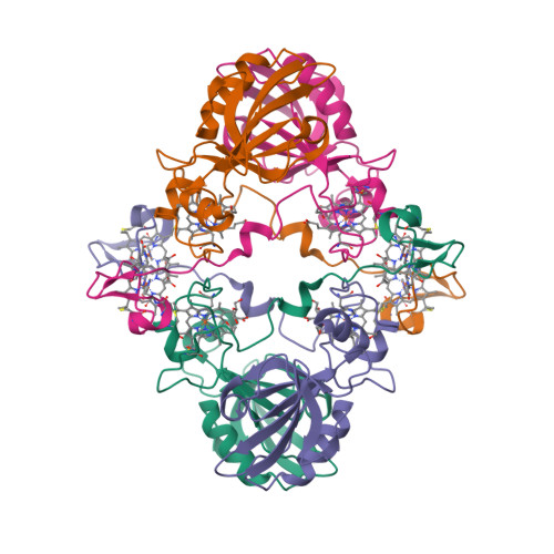
Octahaem sulphite reductase MccA

The epsilonproteobacterium Wolinella succinogenes is able to grow by sulphite respiration with formate as electron donor [1], thanks to the octahaem cytochrome c MccA that catalyses the six-electron reduction of sulphite to sulphide:

HSO3 + 6 H+ + 6 e → HS + 3 H2O

The crystal structure of MccA has been determined at 2.2 Å resolution [2, 3]. The enzyme exists as a homotrimer showing a novel fold and haem arrangement. The heterobimetallic active centre contains a Cu(I) ion and a haem c with a Fe—Cu distance of 4.4 Å [4].

a, W. succinogenes MccA binds its substrate sulfite in the dehydrated form, SO2, at the distal axial position of haem 2. At 3.2 Å distance from the sulphur atom, a Cu(I) ion is nearly linearly coordinated by residues C399 and C495.

b, In respiratory haem–copper oxidases, CuB is a redox-active species liganded by three histidine residues and juxtaposed to a haem a3 moiety. The arrangement, with a Fe–Cu distance of 4.9 Å, is optimized to bind O2 and peroxide in a bridging fashion (PDB:3ABM).

  1. Kern, M., Klotz, M.G. and Simon, J. (2011) The Wolinella succinogenes mcc gene cluster encodes an unconventional respiratory sulphite reduction system. Molecular Microbiology 82, 1515—1530.
  2. PDB:4RKM
  3. PDB:4RKN
  4. Hermann, B., Kern, M., La Pietra, L., Simon, J., Einsle, O. (2015) The octahaem MccA is a haem c-copper sulfite reductase. Nature 520, 706—709.

Saturday, February 27, 2016

First eukaryotic photosystem II solved at 2.76 Å

The water-splitting reaction of photosynthesis is catalysed by photosystem II (PSII), a large protein complex located in thylakoid membranes of organisms ranging from cyanobacteria to higher plants [1]. During the last 15 years, a number of crystal structures of PSII from cyanobacteria have been solved. However, no structures of PSII from eukaryots have been reported until now, partly due to the instability of eukaryotic PSII upon isolation. Ago et al. [2] solved the structure of PSII from a red alga Cyanidium caldarium at 2.76 Å resolution [3]. This PSII contains four extrinsic proteins, including the three subunits found in cyanobacterial PSII and the fourth subunit PsbQ' homologous to the PsbQ protein of green algae and higher plants. Furthermore, two novel trans-membrane helices were found in the algal PSII which are not present in cyanobacterial PSII.

  1. Shen, J.-R. (2015) The structure of photosystem II and the mechanism of water oxidation in photosynthesis. Annual Review of Plant Biology 66, 23—48.
  2. Ago, H., Adachi, H., Umena, Y., Tashiro, T., Kawakami, K., Kamiya, N., Tian, L., Han, G., Kuang, T., Liu, Z., Wang, F., Zou, H., Enami, I., Miyano, M. and Shen, J.-R. (2016) Novel features of eukaryotic photosystem II revealed by its crystal structure analysis from a red alga. J. Biol. Chem. 291, 5676—5687.
  3. PDB:4YUU

Saturday, January 30, 2016

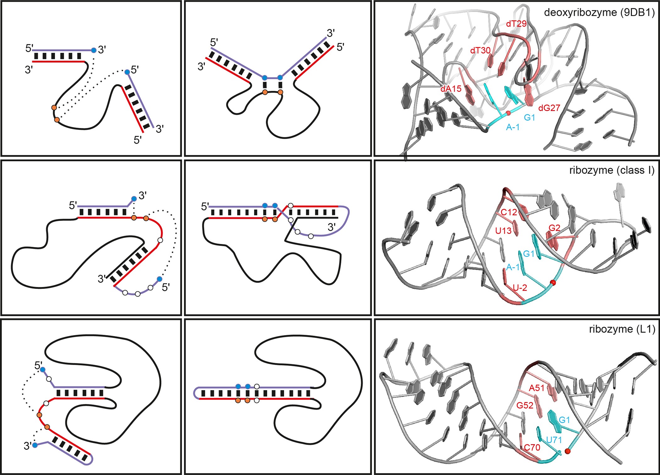
Crystal structure of the DNAzyme 9DB1

The first ever crystal structure of a deoxyribozyme has been solved at 2.8 Å resolution [1—3]. The work by researchers from Max Planck Institute for Biophysical Chemistry (Göttingen, Germany) also sheds light on a difference in catalytic mechanism of ribozymes and deoxyribozymes [4]:

Ribozymes use RNA’s 2´-hydroxyl groups, which are absent in DNA, for structural interactions or directly for catalysis. The new structure shows why the lack of these groups doesn’t diminish the catalytic activity of DNAzymes. The missing hydroxyls make DNA’s sugar-phosphate backbone more flexible, allowing acrobatic conformations that compensate for the absent hydroxyls in DNAzymes.

  1. Ponce-Salvatierra, A., Wawrzyniak-Turek, K., Steuerwald, U., Höbartner, C. and Pena, V. (2016) Crystal structure of a DNA catalyst. Nature 529, 231—234.
  2. PDB:5CKK
  3. PDB:5CKI
  4. Borman, S. (2016) After two decades of trying, scientists report first crystal structure of a DNAzyme. Chemical & Engineering News 94, issue 2, p. 3.

Friday, December 25, 2015

Human Apaf-1 apoptosome at 3.8 Å

The apoptotic protease-activating factor 1 (Apaf-1) exists in normal cells as an autoinhibited monomer. Upon binding to cytochrome c and dATP, Apaf-1 forms a heptameric complex known as the apoptosome. Zhou et al. report an atomic structure of an intact human Apaf-1 apoptosome at 3.8 Å resolution determined by single-particle, cryo-electron microscopy [1, 2].

  1. Zhou, M., Li, Y., Hu, Q., Bai, X.-c., Huang, W., Yan, C., Scheres, S.H.W. and Shi, Y. (2015) Atomic structure of the apoptosome: mechanism of cytochrome c- and dATP-mediated activation of Apaf-1. Genes & Development 29, 2349—2361.
  2. PDB:3JBT

Thursday, November 19, 2015

A magnetic protein biocompass?

A team of scientists from Peking University report a putative magnetic receptor (MagR) protein in Drosophila, CG8198 [1]. MagR binds an iron—sulphur cluster and interacts with photoreceptor cryptochrome (Cry) proteins to form a multimeric magnetosensing rod-like complex. Assemblies of these rods were observed orienting themselves in a weak magnetic field. Qin et al. speculate that these structures may function like compasses in living organisms, although the mechanism of magnetoreception in vivo remains a mystery [2].

A complete Cry/MagR magnetosensor protein complex structure model with 10 Crys helically binding to the rod-like MagR polymer consisting of 20 MagRs.
  1. Qin, S., Yin, H., Yang, C., Dou, Y., Liu, Z., Zhang, P., Yu, H., Huang, Y., Feng, J., Hao, J., Hao, J., Deng, L., Yan, X., Dong, X., Zhao, Z., Jiang, T., Wang, H.-W., Luo, S.-J. and Xie, C. (2016) A magnetic protein biocompass. Nature Materials 15, 217–226.
  2. Cyranoski, D. (2015) Long-sought biological compass discovered: Protein complex offers explanation for how animals sense Earth’s magnetic pull. Nature 527, 283–284.

Sunday, September 27, 2015

Nitrite binding modes in CuNIR

Fukuda and Inoue [1] have determined the crystal structure of the C135A mutant of thermostable copper nitrite reductase (CuNIR) from Geobacillus thermodenitrificans in complex with nitrite to 1.55 Å resolution. Interestingly, this high-temperature (320 K) structure [2] displays a near-bidentate binding mode of nitrite distinct from a monodentate mode in a cryogenic structure [3]:

To our knowledge, this is the first case in which the difference in substrate binding modes between cryogenic and high-temperature structures has been visualized by crystallography.

The copper site geometries are given in Table 1.

Table 1 (adapted from [4])

Cu—Ligand Distances (Å) 3X1N (320 K) 3WKP (100 K)
I. Type 1 Cu—residue distances
T1Cu—H95Nδ1 2.08 2.14
T1Cu—H143Nδ1 2.01 1.96
T1Cu—M148Sδ 2.13 2.07
II. Type 2 Cu—residue distances
T2Cu—H100Nε2 2.07 1.96
T2Cu—H134Nε2 2.00 1.95
T2Cu—H294Nε2 1.98 2.01
T2Cu—water 2.02 n/a
III. Type 2 Cu—nitrite distances
T2Cu—Oproximal 2.13 1.97
T2Cu—N 2.21 2.85
T2Cu—Odistal 2.52 3.41
  1. Fukuda, Y. and Inoue, T. (2015) High-temperature and high-resolution crystallography of thermostable copper nitrite reductase. Chemical Communications 51, 6532—6535.
  2. PDB:3X1N
  3. PDB:3WKP
  4. Fukuda, Y. and Inoue, T. (2015) High-temperature and high-resolution crystallography of thermostable copper nitrite reductase. Electronic Supplementary Material.

Tuesday, August 11, 2015

Colourful Compound Interest

I discovered Andy Brunning’s Compound Interest last year and got absolutely hooked on it – and I don’t even teach chemistry! If, perchance, you do teach chemistry and don’t yet know what CI is all about, then you probably should check it out. (And if you want to use the material in the classroom, you can download the high-resolution PDF files.) The topics range from general chemistry to material science, chemical warfare and everyday compounds. You’ve got answers to many questions you always wanted to ask but never had time to find out for yourself, like, “is it worth (not) to refrigerate tomatoes?”. The Undeserved Reputations section is a perfect antidote to the “oh my God, our food is still full of chemicals” stream of rubbish published by your Facebook friends.

Here are ten the top ten some of my CI favourites.

    Metal Ion Flame Test Colours Chart

  1. This is how (I’d like to think) I’ve got interested in chemistry. We used to have a gas hob in our kitchen. I loved the fact that the flame was blue. One day, my brother told me that you can make the flame bright orangey-yellow if you sprinkle it with table salt or bicarbonate of soda. “Why?”, I asked. “Sodium”, was the answer. Unsatisfactory as it was, it stayed in my memory. Yes, chemistry won’t be of any interest to me if not for flame and colours.
  2. Colours of Transition Metal Ions in Aqueous Solution

  3. When they are not busy burning or, better still, exploding stuff, your archetypal chemists are often imagined (and therefore portrayed; or is it the other way round?) as hiding behind the test tubes filled with colourful solutions. Which is just as well. The test tubes filled with colourless solutions would be really boring.
  4. What Causes the Colour of Gemstones?

  5. Who didn’t dream of finding a treasure, that is, a pirate’s chest filled with gold and jewels? Wait. I still dream of that. I remember how surprised I was when, back in elementary school, I read in some book that ruby and sapphire are basically the same mineral corundum, the only difference is in a type of impurity. Well it’s quite an important difference then. Without impurities, most gemstones would be colourless.
  6. The Chemistry of The Colours of Blood

  7. My interest in bioinorganic chemistry (even though at the time I didn’t know at it was called that) was also awakened in school, when I learned that some animals have blue blood. I also discovered that, contrary to what anatomy textbooks show, veins do not carry blue blood in humans. I am not sure if I was relieved or disappointed. Later, already in the university, I read about a Soviet-developed fluorocarbon-based blood substitute nicknamed “Blue Blood”. Fascinating stuff.
  8. The Chemicals Behind the Colours of Autumn Leaves

  9. I remember, as a child, reading, or rather browsing, an illustrated book about plants (translated from English), with many beautiful colour photographs. “This apple is yellow because of anthocyanin”. Next page: “This apple is yellow because of carotene”. Next page: “This apple is green because of chlorophyll”. The realisation dawned that, apple-wise, being green is not only necessary but sometimes sufficient.

    But what about leaves? When autumn comes, chlorophyll starts to break down and we get to see other pigments in them. Apart from caroteinoids and flavonoids, there are also coloured chlorophyll degradation products, termed “rusty pigments”.

  10. The Chemistry of Stain Removal

  11. Sometimes, however, we want to get rid of all these beautiful colours. The infographic shows the chemical methods of achieving that, although I am not sure that “enzymatic stains” is a correct name for stains caused by blood or grass (yes, haem and chlorophyll again!).
  12. The Atmospheres of the Solar System

  13. Alchemists associated seven metals with seven planets (which included the sun and the moon). At the time, it seemed to be quite reasonable. Now that nobody expects Mercury to be made of mercury (and, for that matter, Pluto to be made of plutonium), precious little is known about composition of these planets. About their atmospheres, we’ve learned a bit more. Hey, isn’t it amazing that Mercury’s atmosphere has by far highest percentage of molecular oxygen (42%) compared to any other atmosphere in Solar system? We still won’t be able to breathe there though, because its atmosphere is way too thin (its surface pressure is less than 10−14 bar).
  14. The Metals in UK Coins

  15. Compared to gemstones, coins are so much duller, especially now that we don’t come across either gold or silver coins any longer. Continuing the alchemical tradition, we can say that modern British coins of 20 pence and higher are mostly from Venus (that is, copper), while 1 p, 2 p, 5 p and 10 p coins are mostly from Mars (i.e. iron). Of course, you can find much more metal variety in commemorative coins.
  16. The Metal Reactivity Series

  17. In contrast to their salts, aqueous complexes and gemstones, pure metals do not offer a great variety of colours. Copper is red, gold is yellow and caesium is yellowish; the rest are coming in many shades of grey. But their chemical behaviour is wildly different, as this infographics shows. You don’t need a sophisticated lab equipment or fancy reagents, just water and some (diluted) acids. If there’s no reaction whatsoever, you’ve got a precious metal. Easy!
  18. Analytical Chemistry – Infrared (IR) Spectroscopy

  19. Did I tell you that my first love, as far as the world of analytical chemistry is concerned, was vibrational spectroscopy? If not, I’m telling you now. I’ve never got to do any experiment worthy of a publication, because if I did, believe me, it would have been awesome. This infographics reminded me of happy days of my studenthood when I knew and cared more about amide bands (bless them) than about money or my future career.

Wednesday, June 10, 2015

There is no perfect language

From The Information: A History, A Theory, A Flood by James Gleick:

It was once thought that a perfect language should have an exact one-to-one correspondence between words and their meanings. There should be no ambiguity, no vagueness, no confusion. Our earthly Babel is a falling off from the lost speech of Eden: a catastrophe and a punishment. “I imagine,” writes the novelist Dexter Palmer, “that the entries of the dictionary that lies on the desk in God’s study must have one-to-one correspondences between the words and their definitions, so that when God sends directives to his angels, they are completely free from ambiguity. Each sentence that He speaks or writes must be perfect, and therefore a miracle.” We know better now. With or without God, there is no perfect language.

Leibniz thought that if natural language could not be perfect, at least the calculus could: a language of symbols rigorously assigned. “All human thoughts might be entirely resolvable into a small number of thoughts considered as primitive.” These could then be combined and dissected mechanically, as it were. “Once this had been done, whoever uses such characters would either never make an error, or, at least, would have the possibility of immediately recognizing his mistakes, by using the simplest of tests.” Gödel ended that dream.

On the contrary, the idea of perfection is contrary to the nature of language. Information theory has helped us understand that — or, if you are a pessimist, forced us to understand it.

Monday, June 01, 2015

Periodic Videos

It’s been a while since I posted anything on this blog, but now I’m back.

This is a very cool collection of videos, “a lesson about every single element on the periodic table”. Featuring Professor and a really awesome reaction, here’s one about one of my favourite elements. Yes, iron is in my blood! (In yours too.)

Wednesday, October 15, 2014

Metals

From A Dictionary of Symbols by Juan Eduardo Cirlot (translated by Jack Sage):
In astrology they are called ‘terrestrial’ or ‘subterranean planets’, because of the analogous correspondences between the planets and the metals. For this reason astrologers consider that there are only seven metals (influenced by the same number of spheres), which does not mean that mankind during the astrobiological period did not recognize more. As Piobb has pointed out, some engineers have noted that the seven planetary metals make up a series which is applicable to the system of the twelve polygons. But, apart from the theory of correspondences, the metals symbolize cosmic energy in solidified form and, in consequence, the libido. On this basis, Jung has asserted that the base metals are the desires and the lusts of the flesh. Extracting the quintessence from these metals, or transmuting them into higher metals, is equivalent to setting creative energy free from the fetters of the sense world, a process identical with what esoteric tradition and astrology regard as liberation from the ‘planetary influences’. The metals can be grouped within a progressive ‘series’ in which each metal displays its hierarchical superiority over the one preceding it, with gold as the culminating point of the progression. This is why, in certain rites, the neophyte is required to divest himself of his ‘metals’ — coins, keys, trinkets — because they are symbolic of his habits, prejudices and characteristics, etc. We, for our part, however, are inclined to believe that in each particular pairing of planet with metal (as Mars with iron) there is an essential element of the ambitendent, in that its positive quality tends one way and its negative defect tends the other. Molten metal is an alchemic symbol expressing the coniunctio oppositorum (the conjunction of fire and water), related to mercury, Mercury and Plato’s primordial, androgynous being. And at the same time, the solid or ‘closed’ properties of matter emphasize its symbolism as a liberator — hence the connexion with Hermes the psychopomp <...> . The correspondences between the planets and the metals, from inferior to superior, are: Saturn — lead, Jupiter — tin, Mars — iron, Venus — copper, Mercury — mercury, Moon — silver, Sun — gold.

Tuesday, September 23, 2014

Pseudomonas fluorescens PhoX

Alkaline phosphatases (EC 3.1.3.1) occur widely in nature and are found in all three domains of life [1]. The Escherichia coli PhoA enzyme has been extensively studied whereas PhoX family of alkaline phosphatases are only minimally characterised and show no sequence similarity to other phosphotransfer enzymes. Yong et al. [2] determined high-resolution crystal structures for native PhoX from Pseudomonas fluorescens [3] and for its complexes with phosphate [4], a nonhydrolysable ATP analogue adenosine-5′-[β,γ-methylene]triphosphate (AMP-PCP) [5], and the putative transition-state mimic vanadate [6]. The active site contains two antiferromagnetically coupled ferric ions (Fe3+), three calcium ions (Ca2+), and an oxo group bridging one Ca2+ and two Fe3+ ions.

Cartoon representation of P. fluorescens PhoX crystal structure.
The PhoX active site containing bound phosphate [1, Fig. 2c].
A model for the catalytic mechanism of PhoX [1, Fig. 3d].
The transition state is indicated with the double dagger (‡) symbol.
  1. Millán, J.L. (2006) Alkaline Phosphatases: Structure, substrate specificity and functional relatedness to other members of a large superfamily of enzymes. Purinergic Signalling 2, 335–341.
  2. Yong, S.C., Roversi, P., Lillington, J., Rodriguez, F., Krehenbrink, M., Zeldin, O.B., Garman, E.F., Lea, S.M. and Berks, B.C. (2014) A complex iron-calcium cofactor catalyzing phosphotransfer chemistry. Science 345, 1170—1173.
  3. PDB:4A9V
  4. PDB:4ALF
  5. PDB:4AMF
  6. PDB:3ZWU

Tuesday, August 26, 2014

Ogres are not like cakes

I was intrigued by the article in New Scientist which starts with the question, “Do you speak chemistry?” [1]. So much that I asked my friend to send me the original paper [2] authored by the Bartosz Grzybowski group of Northwestern University in Evanston, Illinois. It is a curious reading.

Don’t get me wrong. I have nothing against the analogies. I love the analogies. If the linguistic analogy works for chemistry, it’s fine by me. As long as everybody understands that it is just an analogy.

The authors try to “demonstrate that a natural language such as English and organic chemistry have the same structure in terms of the frequency of, respectively, text fragments and molecular fragments”. How do they do that? They start by looking at the maximum common substrings (MCS) found in 100 sentences randomly chosen from English Wikipedia.

Perhaps not surprisingly, the most common fragment of the sentences is “e”, followed by “a” and “o”.
That is surprising to me though, considering that only “a” is a word in English. I wouldn’t be surprised if it happened to be Spanish Wikipedia. Are the authors talking about letter frequency per chance? But the “top three” letters in English (from most to least common) are known to be E, T, A while in Spanish they are E, A, O. Anyway, they show that the distribution of the fragments, whatever they are, follows the power law. Then they show that the distribution of the common molecular fragments, derived from the corpus of organic molecules, also follows the power law. Big deal: so do the earthquake magnitudes, populations of cities and stock market crashes [3]. Cadeddu et al. do not seem to be bothered with that at all:
We have just shown that there exists a set of molecular fragments with which organic molecules can be described akin to a language.
So far so bad; whether you are a linguist, a computational chemist or an organic chemist, both methodology and conclusions of this paper are bound to make you cringe. So, my immediate reaction was to dismiss it altogether. Ogres are not like cakes. Organic molecules are not like a language. End of story.

But could it be that I am missing something? On the one hand, the language of chemistry — whether we are talking trivial names, systematic names, or graphical diagrams — is very much like any other language: a system of communication. On the other hand, the molecules themselves are not. Unless they are the information macromolecules. The message encoded in a single DNA molecule can be very much abstracted from its chemical structure. Without any doubt, genetic code is a communication system, therefore it is a language, although not man-made.

It’s interesting that the authors view organic molecules as “sentences” rather than “words”; the latter would be the nomenclaturist’s approach. I guess it depends on your taste, or language preferences. Most systematic chemical names look alien in English but would fit rather nicely in German or Finnish. I personally view any chemical name as a noun phrase describing a corresponding molecular entity; a molecular entity itself is not a noun phrase. However, in natural languages, there rarely is a confusion regarding the boundaries of a word:

a word is the smallest element that may be uttered in isolation with semantic or pragmatic content (with literal or practical meaning).
On the contrary, Grzybowski’s “words” are the molecular fragments which do not exist in isolation. It is also worth noting that in the world of biopolymers, say nucleic acids, each monomer (as complex as any of Grzybowski’s “sentences”), is often represented as a letter, while an entire bacterial genome (still a single DNA molecule) could be considered a War and Peace (or Crime and Punishment).

Cadeddu et al. further claim that linguistic approach identifies the symmetry/repeat units in molecules such as α-cyclodextrin and porphyrin:

We emphasize that this is not a small feat given we have not even considered any (x, y, z) coordinates of the atoms making up these molecules and performed no linear-algebra analyses to find symmetries which, incidentally, can be a computationally intensive procedure involving manipulation of matrices.
I find this modest remark regarding the size of the “feat” within the body of a scientific article in a respectable journal really cute. Are the authors even aware that there are chemical similarity/substructure search engines? You don’t need atomic coordinates to identify the fragments with the same connectivity.

Which brings me to the final point. What is the “chemical linguistics” anyway? If the “words” of chemistry, as postulated in [2], are nothing else but molecular fragments, or substructures, then the chemoinformaticians were doing the substructure search of chemical databases for donkey’s years without knowing that it is called chemical linguistics. I am aware of completely different use of this term in a sense “mining of natural language texts for chemical information” [4, 5]. This latter use is well-established and I think applying the name “chemical linguistics” to unrelated area will only confuse everybody.

  1. Aron, J. (2014) Language of chemistry is unveiled by molecular make-up. New Scientist no. 2981, p. 8.
  2. Cadeddu, A., Wylie, E.K., Jurczak, J., Wampler-Doty, M. and Grzybowski, B.A. (2014) Organic chemistry as a language and the implications of chemical linguistics for structural and retrosynthetic analyses. Angewandte Chemie 126, 8246—8250.
  3. Buchanan, M. (2000) Ubiquity, Weidenfeld & Nicolson, London.
  4. Goebels, L., Grotz, H., Lawson, A.L., Roller, S. and Wisniewski, J. (2005) Method and software for extracting chemical data. Patent DE 102005020083 A1.
  5. Day, N.E., Corbett, P.T. and Murray-Rust, P. (2007) Semantic chemical publishing. ACS National Meeting #233, Chicago.

Thursday, July 31, 2014

F420-reducing [NiFe]-hydrogenase at 1.7 Å

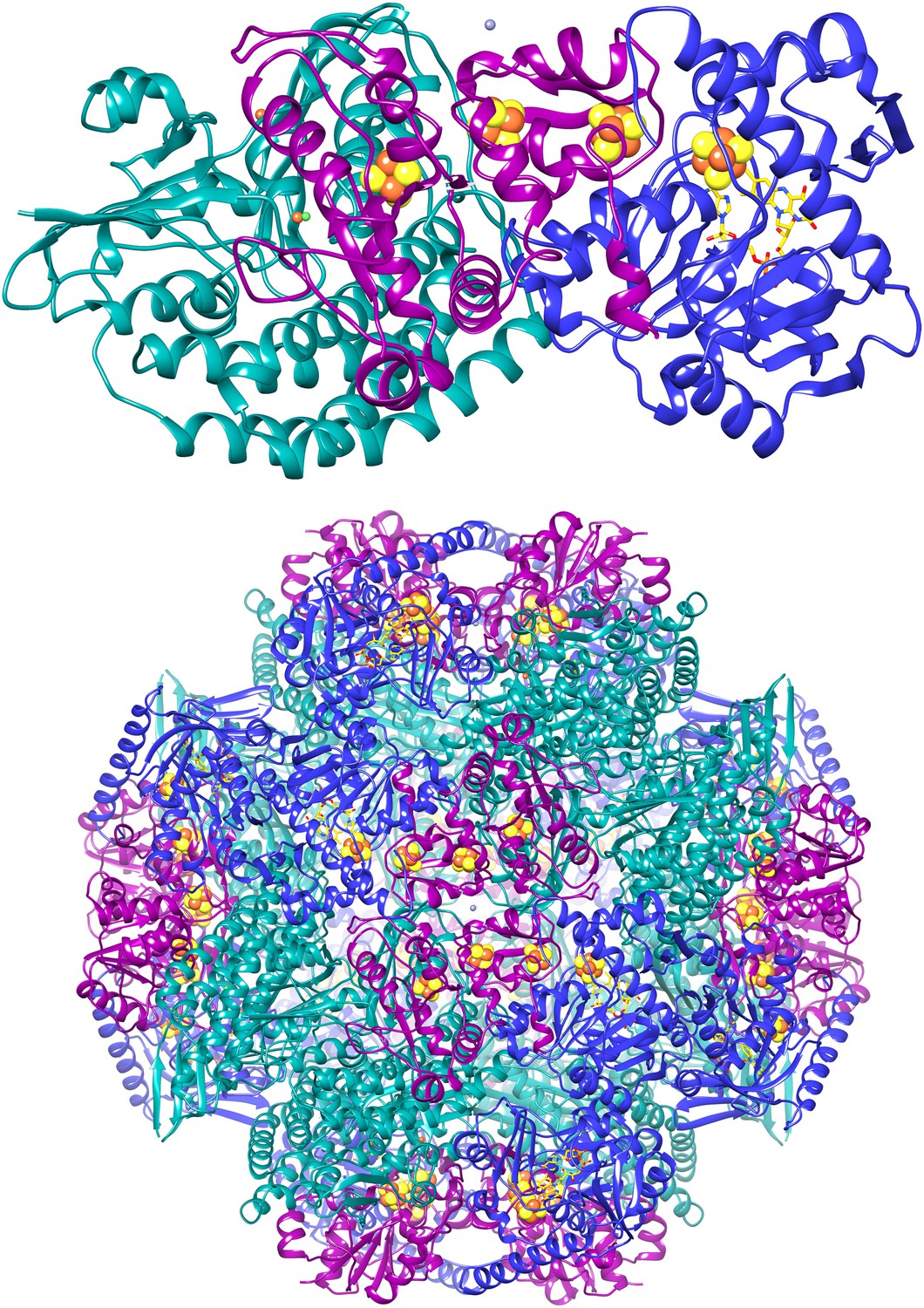
The F420-reducing [NiFe]-hydrogenase (FrhABG; EC 1.12.98.1) catalyses the reversible redox reaction between coenzyme F420 and H2. FrhABG is a group 3 [NiFe]-hydrogenase with a dodecameric quaternary structure recently revealed by high-resolution cryo-electron microscopy [1]. Vitt et al. report the crystal structure of FrhABG from Methanothermobacter marburgensis at 1.7 Å resolution [2, 3] and compare it with the structures of group 1 [NiFe]-hydrogenases, the only previously structurally characterised group.

  1. Allegretti, M., Mills, D.J., McMullan, G., Kühlbrandt, W. and Vonck, J. (2014) Atomic model of the F420-reducing [NiFe] hydrogenase by electron cryo-microscopy using a direct electron detector. eLife 3, e01963.
  2. Vitt, S., Ma, K., Warkentin, E., Moll, J., Pierik, A.J., Shima, S. and Ermler, U. (2014) The F420-reducing [NiFe]-hydrogenase complex from Methanothermobacter marburgensis, the first X-ray structure of a group 3 family member. J. Mol. Biol. 426, 2813—2826.
  3. PDB:4OMF

Monday, June 30, 2014

Phycocyanin against Alzheimer’s?

Could a light-harvesting protein phycocyanin be used as a novel drug against Alzheimer’s disease (AD) [1, 2]?

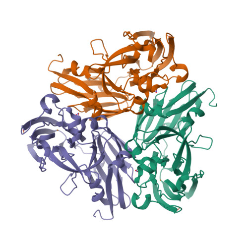
In the present study, intact hexameric phycocyanin was isolated and crystallized from the cyanobacterium Leptolyngbya sp. N62DM, and the structure was solved to a resolution of 2.6 Å. Molecular docking studies show that the phycocyanin αβ-dimer interacts with the enzyme β-secretase, which catalyzes the proteolysis of the amyloid precursor protein to form plaques. The molecular docking studies suggest that the interaction between phycocyanin and β-secretase is energetically more favorable than previously reported inhibitor-β-secretase interactions. Transgenic Caenorhabditis elegans worms, with a genotype to serve as an AD-model, were significantly protected by phycocyanin. Therefore, the present study provides a novel structure-based molecular mechanism of phycocyanin-mediated therapy against AD.
  1. Singh, N.K., Hasan, S.S., Kumar, J., Raj, I., Pathan, A.A., Parmar, A., Shakil, S., Gourinath, S. and Madamwar D. (2014) Crystal structure and interaction of phycocyanin with β-secretase: A putative therapy for Alzheimer's disease. CNS Neurol. Disord. Drug Targets 13, 691—698.
  2. PDB:4L1E

Saturday, May 24, 2014

[Fe3S4] ferredoxin from Rhodopseudomonas palustris

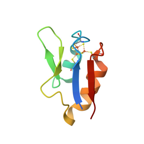
The crystal structure of a novel [Fe3S4] ferredoxin associated with CYP194A4 from Rhodopseudomonas palustris has been solved at 2.15 Å resolution [1—3]. The ferredoxin, HaPuxC, contains an atypical CXXHXXC(X)nCP iron-sulphur cluster-binding motif. HaPuxC is the first P450 electron-transfer partner of this type to be structurally characterised.

  1. Zhang, T., Zhang, A., Bell, S.G., Wong, L.-L. and Zhou, W. (2014) The structure of a novel electron-transfer ferredoxin from Rhodopseudomonas palustris HaA2 which contains a histidine residue in its iron-sulfur cluster-binding motif. Acta Crystallographica D70, 1453—1464.
  2. PDB:4ID8
  3. PDB:4OV1

Thursday, May 01, 2014

Tetracalcium octachromium(3+) strontium octacarbonate hexadecahydroxide sulfate pentaicosahydrate

The Polar Bear peninsula in Western Australia is one of the many places on this planet I never heard before. The reason I mention it now is that a new mineral named putnisite was discovered there, and this mineral caused a bit of a stir recently, for being “completely unique and unrelated to anything”. In fact, if you Google “Polar Bear peninsula”, all you find is putnisite.

In 2007, specimens of an unknown mineral forming purple crystals (a) were collected at the Polar Bear peninsula while prospecting for nickel and gold. The specimens were eventually forwarded to Peter Elliott, a research associate with the South Australian Museum, for examination.

(a)

Elliott et al. [2] report the composition and crystal structure of this unique mineral, named in honour of mineralogists Christine and Andrew Putnis of the Institut für Mineralogie, Universtität Münster, Germany. The compositional name for putnisite I come up with is “tetracalcium octachromium(3+) strontium octacarbonate hexadecahydroxide sulfate pentaicosahydrate”. Curiously, Mindat and Mineralienatlas give the molecular formula containing only 23 molecules of water.

(b)

The crystal structure (b) was determined from single-crystal X-ray diffraction data. Cr(OH)4O2 octahedra (red) link by edge-sharing to form an eight-membered ring. At the centre of each ring lies a decacoordinated Sr2+ cation (purple). The rings are decorated by carbonate triangles (green), each of which links by corner-sharing to two Cr(OH)4O2 octahedra. Rings are linked by Ca(H2O)4O4 polyhedra (blue) to form a sheet parallel to the (100) plane. Adjacent sheets are joined along the [100] direction by corner-sharing sulfate tetrahedra (yellow).

  1. Mills, R. (2014) New mineral shows nature’s infinite variability. Phys.org.
  2. Elliott, P., Giester, G., Rowe, R. and Pring, A. (2014) Putnisite, SrCa4Cr3+8(CO3)8SO4(OH)16·25H2O, a new mineral from Western Australia: description and crystal structure. Mineralogical Magazine 78, 131—144.

Tuesday, April 22, 2014

Thermochromatium tepidum LH1—RC complex at 3.0 Å

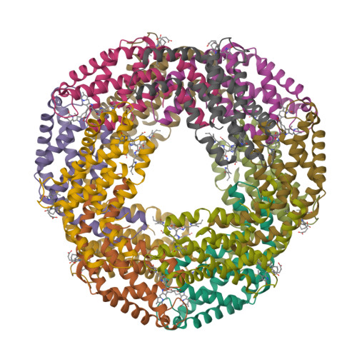
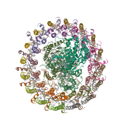
The light-harvesting core antenna (LH1) and the reaction centre (RC) of purple photosynthetic bacteria form a supramolecular complex (LH1—RC) to use sunlight energy in a highly efficient manner. Niwa et al. [1—4] report the first near-atomic structure of a LH1—RC complex, namely that of a Ca2+-bound complex from Thermochromatium tepidum. The RC is surrounded by 16 heterodimers of the LH1 αβ-subunit that form a completely closed structure. The Ca2+ ions are located at the periplasmic side of LH1. Thirty-two bacteriochlorophyll a and sixteen spirilloxanthin molecules in the LH1 ring form an elliptical assembly.

  1. Niwa, S., Yu, L.-J., Takeda, K., Hirano, Y., Kawakami, T., Wang-Otomo, Z.-Y. and Miki, K. (2014) Structure of the LH1—RC complex from Thermochromatium tepidum at 3.0 Å. Nature 508, 228—232.
  2. PDB:3WMM
  3. PDB:4V8K

Thursday, February 27, 2014

Smell of pine vs climate change

That’s right: the smell of pine trees from boreal forests could mitigate the global warming — provided that the global warming doesn’t kill the forests first [1]. The volatile organic compounds (VOCs), responsible for the smell of pine, react with atmospheric oxygen to form aerosols. These aerosols scatter solar radiation and also act as cloud condensation nuclei, thereby affecting the Earth’s radiation balance. An international group including researchers from Finland, Germany, Denmark and USA has discovered a direct pathway leading from several biogenic VOCs, such as monoterpenes, to the formation of extremely low-volatility vapours (ELVOCs) [2].

These vapours form at significant mass yield in the gas phase and condense irreversibly onto aerosol surfaces to produce secondary organic aerosol, helping to explain the discrepancy between the observed atmospheric burden of secondary organic aerosol and that reported by many model studies. We further demonstrate how these low-volatility vapours can enhance, or even dominate, the formation and growth of aerosol particles over forested regions, providing a missing link between biogenic VOCs and their conversion to aerosol particles.

Structures of the main VOCs studied by Ehn et al. [2]

The air was sampled in Hyytiälä, Finland and the chamber experiments were conducted at the Jülich Research Centre in Germany.

  1. McGrath, M. Smell of forest pine can limit climate change. BBC News, 26 February 2014.
  2. Ehn, M., Thornton, J.A., Kleist, E., Sipilä, M., Junninen, J., Pullinen, I., Springer, M., Rubach, F., Tillmann, R., Lee, B., Lopez-Hilfiker, F., Andres, S., Acir, I.-H., Rissanen, M., Jokinen, T., Schobesberger, S., Kangasluoma, J., Kontkanen, J., Nieminen, T., Kurtén, T., Nielsen, L.B., Jørgensen, S., Kjaergaard, H.G., Canagaratna, M., Dal Maso, M.D., Berndt, T., Petäjä, T., Wahner, A., Veli-Matti Kerminen, V.-M., Kulmala, M., Worsnop, D.R., Wildt, J. and Mentel, T.F. (2014) A large source of low-volatility secondary organic aerosol. Nature 506, 476—479.

Saturday, January 18, 2014

Sodium chloride revisited

Everybody knows that the formula of sodium chloride is NaCl. Right? Right. But recently, the team of Artem Oganov at Stony Brook University have shown that there are other stable types of crystalline sodium chloride. They have predicted several thermodynamically stable compounds: Na3Cl, Na2Cl, Na3Cl2, NaCl3, and NaCl7. Moreover, by utilising high-pressure techniques, they synthesised cubic and orthorhombic NaCl3 and two-dimensional tetragonal Na3Cl [1, 2].

NaCl3 (space group Pm3n)
Na3Cl (space group P4/mmm)

“One of these materials — Na3Cl — has a fascinating structure”, says Oganov. “It is comprised of layers of NaCl and layers of pure sodium. The NaCl layers act as insulators; the pure sodium layers conduct electricity” [3].

  1. Zhang, W., Oganov, A.R., Goncharov, A.F., Zhu, Q., Boulfelfel, S.E., Lyakhov, A.O., Stavrou, E., Somayazulu, M., Prakapenka, V.B. and Konôpková, Z. (2013) Unexpected stable stoichiometries of sodium chlorides. Science 342, 1502—1505; arXiv:1310.7674v1
  2. Ibáñez Insa, J. (2013) Reformulating table salt under pressure. Science 342, 1459—1460.
  3. SBU team discovers new compounds that challenge the foundation of chemistry. Stony Brook University Newsroom, December 19, 2013.

Tuesday, November 26, 2013

Magnetochrome-containing iron oxidase MamP

Magnetotactic bacteria (MTB) are a diverse group of prokaryotes that have a singular ability to align with geomagnetic field lines. This ability is due to special organelles called magnetosomes. Magnetosomes are composed of single-magnetic-domain nanocrystals of magnetite [Fe(II)Fe(III)2O4] or greigite [Fe(II)Fe(III)2S4] embedded in biological membrane.

“Magnetochrome” is a name proposed in 2012 by Marina Siponen and co-authors for a cytochrome domain conserved within all known MTB and not found in any other species to date [1]. Recently, the crystal structure of the magnetosome-associated protein MamP has been solved at 1.8 Å resolution [2—4]. The minimal unit of MamP is a dimer. Each monomer consists of a PDZ domain fused to two magnetochrome domains. It was also shown in an in vitro mineralisation experiment that MamP functions as an iron oxidase mediating the iron(III) ferrihydrite production from iron(II) [2]:

4Fe2+ + 7H2O → 2Fe2O3·H2O + 12H+ + 4e
  1. Siponen, M.I., Adryanczyk, G., Ginet, N., Arnoux, P. and Pignol, D. (2012) Magnetochrome: a c-type cytochrome domain specific to magnetotatic bacteria. Biochemical Society Transactions 40, 1319—1323.
  2. Siponen, M.I., Legrand, P., Widdrat, M., Jones, S.R., Zhang, W.-J., Chang, M.C.Y., Faivre, D., Arnoux, P. and Pignol, D. (2013) Structural insight into magnetochrome-mediated magnetite biomineralization. Nature 502, 681—684.
  3. PDB:4JJ0
  4. PDB:4JJ3