Monday, December 24, 2012

Macromolecular Xmas decorations

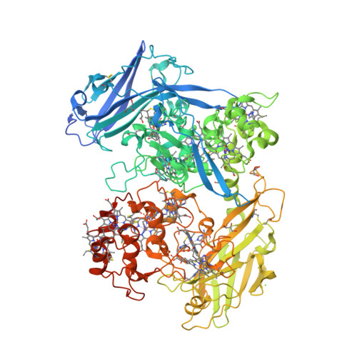
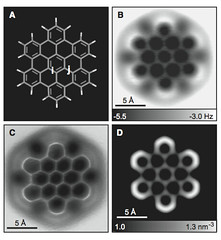
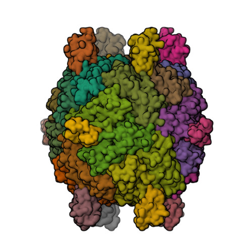
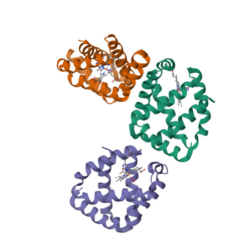
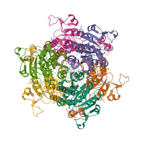
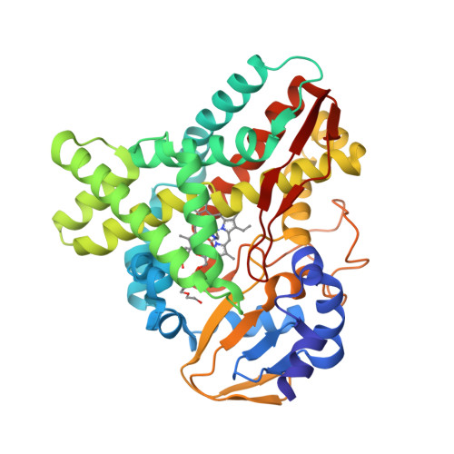
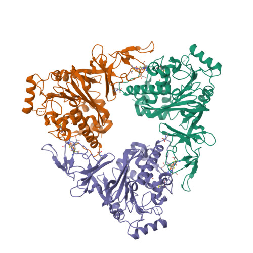
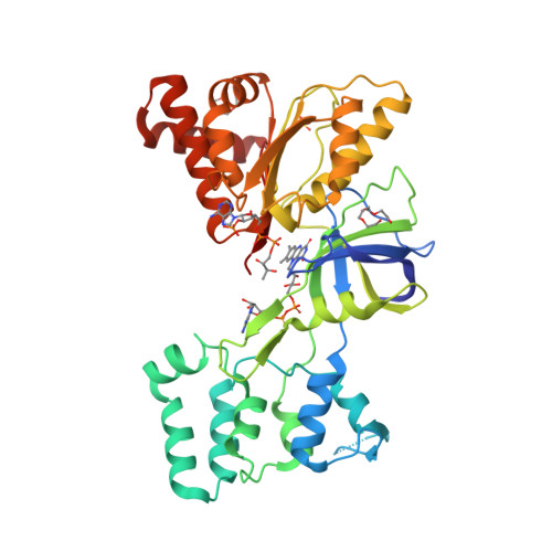
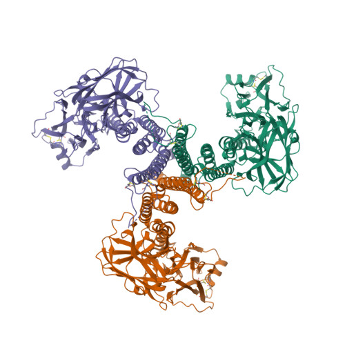
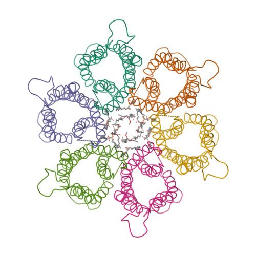
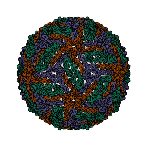
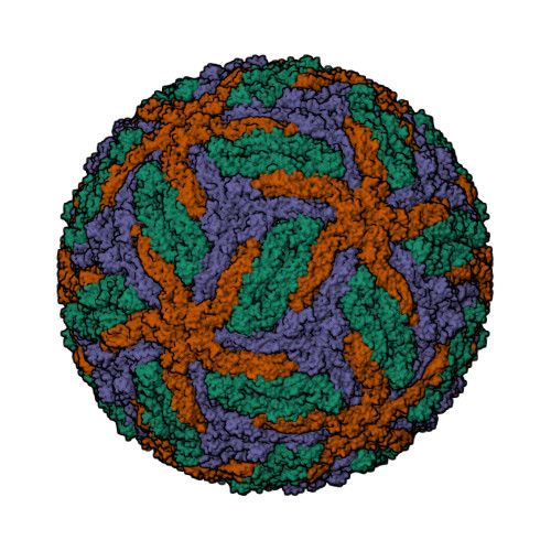
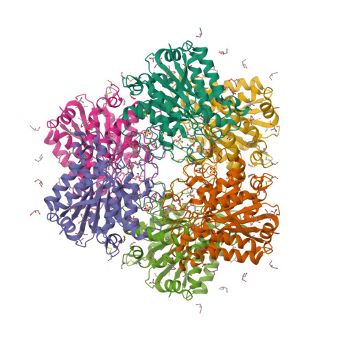
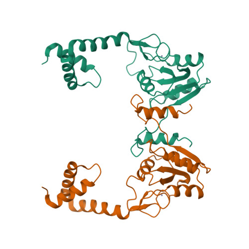
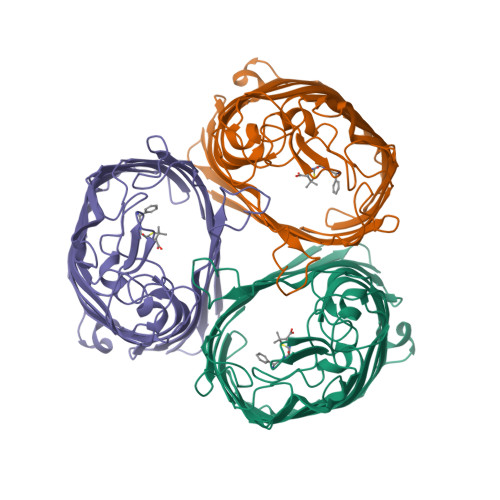
This year I don’t have a Christmas tree. (There aren’t many in Fuerteventura.) But I’ve got some models from this week’s new PDB structures [1—5] which look Christmassy enough to decorate my blog. Merry Christmas and a happy New Year everybody!


  1. Hui, F., Scheib, U., Hu, Y., Sommer, R.J., Aroian, R.V. and Ghosh, P. (2012) Structure and glycolipid binding properties of the nematicidal protein Cry5B. Biochemistry 51, 9911—9921.
  2. Lence, E., Tizón, L., Otero, J.M., Peón, A., Prazeres, V.F.V., Llamas-Saiz, A.L., Fox, G.C., van Raaij, M.J., Lamb, H., Hawkins, A.R. and González-Bello, C. (2013) Mechanistic basis of the inhibition of type II dehydroquinase by (2S)- and (2R)-2-benzyl-3-dehydroquinic acids. ACS Chem. Biol. 8, 568—577.
  3. Strugatsky, D., McNulty, R., Munson, K., Chen, C.-K., Soltis, S.M., Sachs, G. and Luecke, H. (2013) Structure of the proton-gated urea channel from the gastric pathogen Helicobacter pylori. Nature 493, 255—258.
  4. Tang, Q., Gao, P., Liu, Y.-P., Gao, A., An, X.-M., Liu, S., Yan, X.-X. and Liang, D.-C. (2012) RecOR complex including RecR N-N dimer and RecO monomer displays a high affinity for ssDNA. Nucleic Acids Res. 40, 11115—11125.
  5. Ziervogel, B.K. and Roux, B. (2013) The binding of antibiotics in OmpF porin. Structure 21, 76—87.

Tuesday, November 20, 2012

The discovery of the quantum dot

Louis Brus talks about his discovery of colloidal quantum dots in 1980s.

One of the truisms of science is that the basic research scientists who invent something are not the best judges of where it’s useful.

Monday, October 29, 2012

Undecahaem cytochrome from Shewanella

The first crystal structures of the 11-haem cytochrome from Shewanella sp. strain HRCR-6 have been solved [1], both ligand-free [2] and in complex with iron chelates Fe(III)-citrate [3] and Fe(III)-nitrilotriacetate [4]. The authors propose that

the region around heme 7 could be a rudimentary active site for association of soluble organic redox partners, which would be consistent with the UndA functioning as an enzyme with broad, but differential, specificity to a variety of substrates, in contrast to a nonspecific cathode on the cell surface <such as decaheme cytochrome MtrF>.

  1. Edwards, M.J., Hall, A., Shi, L., Fredrickson, J.K., Zachara, J.M., Butt, J.N., Richardson, D.J. and Clarke, T.A. (2012) The crystal structure of the extracellular 11-heme cytochrome UndA reveals a conserved 10-heme motif and defined binding site for soluble iron chelates. Structure 20, 1275—1284.
  2. PDB:3UCP
  3. PDB:3UFH
  4. PDB:3UFK

Friday, September 21, 2012

Visualising hexabenzocoronene

A few years ago, I wrote that we do not know how to draw ferrocene or a nitro group. (Still true.) Is the situation with polycyclic aromatic hydrocarbons any better?

Take hexabenzo[bc,ef,hi,kl,no,qr]coronene, one of the subjects of the single-molecule visualisation study published last week in Science [1]. One way to draw it shown in diagram (a):

(a)

I chose this one (out of many other possible Kekulé representations) because I can reproduce it on a paper napkin (beermat, Post-it note, you name it). If you look carefully, you will notice that the central ring and the six outermost rings are connected with single bonds.

(b)

Continuing the paper-napkin-doodle argument, it is even easier to draw a circle inside of each ring as in all-delocalised representation (b). However, that would not be a preferred diagram from IUPAC point of view [2, GR-6.5]: for example, benzene is acceptable but is preferred. Moreover, “it is generally not acceptable to use curves in two adjacent fused rings”. Still, I’d stick with circles.

The question is, do I have to draw a circle within each ring? Of course not. If I draw seven aromatic rings and connect the with single bonds as shown in (c), the resulting structure will be the same. In this way, I can even save some ink (graphite, chalk, etc.)

(c)

Without the circles, the six rings that surround the central ring in (c) start to look, well, more empty. Using the noncontact atomic force microscopy (NC-AFM), the team behind the study [1] were able to show (and in this case “to show” really means “to show”), that those rings are indeed slightly larger. The C—C bonds in the central ring (i-bonds, 1.417 Å) are 0.03 Å shorter than the bonds connecting that ring with the six outermost rings (j-bonds, 1.447 Å).

  1. Gross, L., Mohn, F., Moll, N., Schuler, B., Criado, A., Guitián, E., Peña, D., Gourdon, A. and Meyer, G. (2012) Bond-order discrimination by atomic force microscopy. Science 337, 1326—1329.
  2. Brecher, J. (2008) Graphical representation standards for chemical structure diagrams (IUPAC Recommendations 2008). Pure Appl. Chem. 80, 277—410.

Thursday, September 06, 2012

IUPAC periodic table?

The cover of the latest issue of Chemistry International features a fragment of Homenatge als elements (Hommage to the Elements) by the Catalan artist Eugènia Balcells. The display in the atrium of the Physics and Chemistry Library at the University of Barcelona takes the shape of the periodic table where each chemical element is represented by its emission spectrum [1]. According to the artist’s website, it “was born as a counterpoint” to the video installation Freqüències (Frequencies).

The Periodic Table Project at the University of Waterloo, Canada is another work of art,

designed by chemistry students from all Canadian provinces and territories, 20 U.S. states, and 14 countries. It can be viewed online and is available as a printed poster.

Also, as a free app for Apple or Android.

Periodic Table Project / Projet Tableau Périodique

Both the Periodic Table Project and Hommage to the Elements use the medium-long form periodic table. The “IUPAC Periodic Table of the Elements” as published at the back of Chemistry International (in this issue, for the first time it includes flerovium and livermorium) has the same shape. Why the quotes? Because, as a matter of fact, there is no such thing as IUPAC-approved periodic table. Jeffery Leigh wrote three years ago that “there is unlikely to be a definitive IUPAC-recommended form of the periodic table” [2]. In my humble opinion, this is unfortunate that IUPAC refuses to take a position on this matter. Eric Scerri takes a view that “IUPAC should in fact take a stance on the membership of particular groups even if this has not been the practice up to this point” [3]. To illustrate this point, he goes to address the Group 3 question. He argues that the most logical composition of this group is Sc, Y, Lu and Lr (rather than Sc, Y, La and Ac), as shown below.

1 2 3 4 5 6 7 8 9 10 11 12 13 14 15 16 17 18
H He
Li Be B C N O F Ne
Na Mg Al Si P S Cl Ar
K Ca Sc Ti V Cr Mn Fe Co Ni Cu Zn Ga Ge As Se Br Kr
Rb Sr Y Zr Nb Mo Tc Ru Rh Pd Ag Cd In Sn Sb Te I Xe
Cs Ba La Ce Pr Nd Pm Sm Eu Gd Tb Dy Ho Er Tm Yb Lu Hf Ta W Re Os Ir Pt Au Hg Tl Pb Bi Po At Rn
Fr Ra Ac Th Pa U Np Pu Am Cm Bk Cf Es Fm Md No Lr Rf Db Sg Bh Hs Mt Ds Rg Cn Nh Fl Mc Lv Ts Og
1 2 3 4 5 6 7 8 9 10 11 12 13 14 15 16 17 18 19 20 21 22 23 24 25 26 27 28 29 30 31 32
In addition to arranging all the elements in a more correct sequence of increasing atomic numbers, the decision to move to a long-form or 32-column table forces the periodic table designer towards just one possible option regarding the question of which elements to place in group 3.

I entirely agree with that. And yet Scerri stops short of proposing that IUPAC should support the 32-column (or “long, long form”, as Leigh put it) periodic table; in fact he explicitly states that he is not suggesting a change of IUPAC policy, viz. that of doing nothing about it. Why? That would be one of the most important and immediately noticeable changes sponsored by IUPAC in decades.

The problem is, sorting out the Group 3 does not resolve the problem how to number the f-block columns. If we stick with 18 groups (blue numbering on the top of the table), that would be really unfair towards the lanthanoids and actinoids. Why don’t we simply number groups from 1 to 32 (red numbers on the bottom of the table)? Sc, Y, Lu and Lr will find themselves in Group 17. So what? It’s not that many people will miss the current Group 17 — nobody really calls these elements anything but “halogens”. And 32 is even more convenient number than 18. I think it’s about time IUPAC took the lead and said how exactly the periodic table should look like.

  1. Alvarez, S. (2012) An artist’s hommage to the elements. Chemistry International 34, 5.
  2. Leigh, J. (2009) Periodic tables and IUPAC. Chemistry International 31, 4—6.
  3. Scerri, E. (2012) Mendeleev’s periodic table is finally completed and what to do about group 3? Chemistry International 34, 28—31.

Tuesday, August 21, 2012

P. aeruginosa bacterioferritin—ferredoxin complex

The X-ray crystal structure of Pseudomonas aeruginosa bacterioferritin (Pa-BfrB) in complex with bacterioferritin-associated ferredoxin (Pa-Bfd) has been solved at 2.0 Å resolution [1, 2].

As the first example of a ferritin-like molecule in complex with a cognate partner, the structure provides unprecedented insight into the complementary interface that enables the [2Fe-2S] cluster of Pa-Bfd to promote heme-mediated electron transfer through the BfrB protein dielectric (~18 Å), a process that is necessary to reduce the core ferric mineral and facilitate mobilization of Fe2+. The Pa-BfrB—Bfd complex also revealed the first structure of a Bfd, thus providing a first view to what appears to be a versatile metal binding domain ubiquitous to the large Fer2_BFD family of proteins and enzymes with diverse functions.
  1. Yao, H., Wang, Y., Lovell, S., Kumar, R., Ruvinsky, A.M., Battaile, K.P., Vakser, I.A. and Rivera, M. (2012) The structure of the BfrB—Bfd complex reveals protein—protein interactions enabling iron release from bacterioferritin. J. Am. Chem. Soc. 134, 13470—13481.
  2. PDB:4E6K

Friday, July 20, 2012

Crystal structure of HGbI

Hell’s Gate globin I from an obligate methanotroph Methylacidiphilum infernorum. Poetry.

  1. Teh, A.-H., Saito, J.A., Baharuddin, A., Tuckerman, J.R., Newhouse, J.S., Kanbe, M., Newhouse, E.I., Rahim, R.A., Favier, F., Didierjean, C., Sousa, E.H.S., Stott, M.B., Dunfield, P.F., Gonzalez, G., Gilles-Gonzalez, M.A., Najimudin, N. and Alam, M. (2011) Hell’s Gate globin I: An acid and thermostable bacterial hemoglobin resembling mammalian neuroglobin. FEBS Lett. 585, 3250—3258.
  2. Pechkova, E., Scudieri, D., Belmonte, L. and Nicolini, C. (2012) Oxygen-bound Hell’s gate globin I by classical versus LB nanotemplate method. J. Cell Biochem. 113, 2543—2548.
  3. PDB:3S1I
  4. PDB:3S1J
  5. PDB:3UBC
  6. PDB:3UBV

Friday, June 15, 2012

Polyoxomolybdate clusters of Mo/W-storage protein

Five years ago, Schemberg et al. reported the crystal structure of molybdenum/tungsten storage protein from Azotobacter vinelandii complexed with polyoxotungstates [1, 2].

Now Kowalewski et al. report the 1.6 Å X-ray structure of the same protein containing a variety of polyoxomolybdate clusters, from Mo3 to Mo8 [3].

Some N2-fixing bacteria prolong the functionality of nitrogenase in molybdenum starvation by a special Mo storage protein (MoSto) that can store more than 100 Mo atoms. The presented 1.6 Å X-ray structure of MoSto from Azotobacter vinelandii reveals various discrete polyoxomolybdate clusters, three covalently and three noncovalently bound Mo8, three Mo5–7, and one Mo3 clusters, and several low occupied, so far undefinable clusters, which are embedded in specific pockets inside a locked cage-shaped (αβ)3 protein complex. <...> The formed polyoxomolybdate clusters of MoSto, not detectable in bulk solvent, are the result of an interplay between self- and protein-driven assembly processes that unite inorganic supramolecular and protein chemistry in a host–guest system.
  1. Schemberg, J., Schneider, K., Demmer, U., Warkentin, E., Müller, A. and Ermler, U. (2007) Towards biological supramolecular chemistry: a variety of pocket-templated, individual metal oxide cluster nucleations in the cavity of a Mo/W-storage protein. Angewandte Chemie International Edition 46, 2408—2413.
  2. PDB:2OGX
  3. Kowalewski, B., Poppe, J., Demmer, U., Warkentin, E., Dierks, T., Ermler, U. and Schneider, K. (2012) Nature’s polyoxometalate chemistry: X-ray structure of the Mo storage protein loaded with discrete polynuclear Mo–O clusters. J. Am. Chem. Soc. 134, 9768—9774.

Thursday, May 10, 2012

P450-flavodoxin fusion enzyme XplA

XplA is a P450-flavodoxin fusion enzyme that mediates the metabolism of the military explosive RDX (1,3,5-trinitro-1,3,5-triazinane) in Rhodococcus rhodochrous 11Y [1]. Bui et al. have conducted a detailed spectroscopic and crystallographic study of this unusual hemoflavoprotein [2, 3].

The XplA P450 has evolved as a reductase (rather than oxidase) of RDX and structural alterations to its heme- and FMN-binding domains have led to reduction potentials for low-spin heme iron Fe3+/Fe2+ and FMNSQ/HQ couples being much more positive than those seen in typical P450s and flavodoxins, but consistent with non-oxidative P450 catalysis. These evolutionary steps have also led to a constricted P450 active site with high affinity for RDX (but also for the small heterocyclic inhibitor imidazole), and also to substantially diminished affinity for FMN in the flavodoxin domain.

  1. Rylott, E.L., Jackson, R.G., Sabbadin, F., Seth-Smith, H.M.B., Edwards, J., Chong, C.S., Strand, S.E., Grogan, G. and Bruce, N.C. (2011) The explosive-degrading cytochrome P450 XplA: biochemistry, structural features and prospects for bioremediation. Biochim. Biophys. Acta 1814, 230—236.
  2. Bui, S.H., McLean, K.J., Cheesman, M.R., Bradley, J.M., Rigby, S.E.J., Levy, C.W., Leys, D. and Munro, A.W. (2012) Unusual spectroscopic and ligand binding properties of the cytochrome P450-flavodoxin fusion enzyme XplA. J. Biol. Chem. 287, 19699—19714.
  3. PDB:4EP6

Tuesday, April 24, 2012

Stachydrine demethylase

Crystal structures were determined for the Rieske-type monooxygenase, stachydrine demethylase, in the unliganded state (at 1.6 Å) and in the product complex (at 2.2 Å) [1—3].

  1. Daughtry, K.D., Xiao, Y., Stoner-Ma, D., Cho, E., Orville, A.M., Liu, P. and Allen, K.N. (2012) Quaternary ammonium oxidative demethylation: X-ray crystallographic, resonance Raman, and UV-visible spectroscopic analysis of a Rieske-type demethylase. J. Am. Chem. Soc. 134, 2823—2834.
  2. PDB:3VCA
  3. PDB:3VCP

Sunday, March 25, 2012

FAD/NADPH-domain of flavocytochrome P450 BM3

The crystal structure of the FAD/NADPH-binding domain of the Bacillus megaterium flavocytochrome P450 BM3 has been solved in both the absence and presence of the ligand NADP+ [1—3].

  1. Joyce, M.G., Ekanem, I.S., Roitel, O., Dunford, A.J., Neeli, R., Girvan, H.M., Baker, G.J., Curtis, R.A., Munro, A.W. and Leys, D. (2012) The crystal structure of the FAD/NADPH-binding domain of flavocytochrome P450 BM3. FEBS J. 279, 1694—1706.
  2. PDB:4DQK
  3. PDB:4DQL

Sunday, February 05, 2012

Carbon—carbon quadruple bond

Quadruple and higher order metal—metal bonds are known for transition metals, lanthanoids and actinoids. But for main group elements? Using four different computational methods, Shaik et al. [1] show that

C2 and its isoelectronic molecules CN+, BN and CB (each having eight valence electrons) are bound by a quadruple bond. The bonding comprises not only one σ- and two π-bonds, but also one weak ‘inverted’ bond, which can be characterized by the interaction of electrons in two outwardly pointing sp hybrid orbitals.
According to Shaik, the existence of the fourth bond in C2 suggests that it is not really diradical C22• [2]:
If C2 were a diradical it would immediately form higher clusters. I think the fact that you can isolate C2 tells you it has a barrier, small as it may be, to prevent that.
  1. Shaik, S., Danovich, D., Wu, W., Su, P., Rzepa, H.S. and Hiberty, P.C. Quadruple bonding in C2 and analogous eight-valence electron species. Nature Chemistry 4, 195—200.
  2. Extance, A. Calculations reveal carbon-carbon quadruple bond. Chemistry World, 29 January 2012.