Showing posts with label biomineralisation. Show all posts

Tuesday, November 26, 2013

Magnetochrome-containing iron oxidase MamP

Magnetotactic bacteria (MTB) are a diverse group of prokaryotes that have a singular ability to align with geomagnetic field lines. This ability is due to special organelles called magnetosomes. Magnetosomes are composed of single-magnetic-domain nanocrystals of magnetite [Fe(II)Fe(III)2O4] or greigite [Fe(II)Fe(III)2S4] embedded in biological membrane.

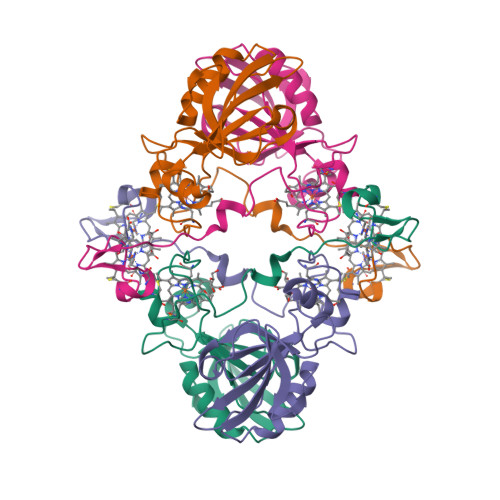
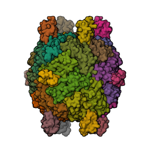
“Magnetochrome” is a name proposed in 2012 by Marina Siponen and co-authors for a cytochrome domain conserved within all known MTB and not found in any other species to date [1]. Recently, the crystal structure of the magnetosome-associated protein MamP has been solved at 1.8 Å resolution [2—4]. The minimal unit of MamP is a dimer. Each monomer consists of a PDZ domain fused to two magnetochrome domains. It was also shown in an in vitro mineralisation experiment that MamP functions as an iron oxidase mediating the iron(III) ferrihydrite production from iron(II) [2]:

4Fe2+ + 7H2O → 2Fe2O3·H2O + 12H+ + 4e
  1. Siponen, M.I., Adryanczyk, G., Ginet, N., Arnoux, P. and Pignol, D. (2012) Magnetochrome: a c-type cytochrome domain specific to magnetotatic bacteria. Biochemical Society Transactions 40, 1319—1323.
  2. Siponen, M.I., Legrand, P., Widdrat, M., Jones, S.R., Zhang, W.-J., Chang, M.C.Y., Faivre, D., Arnoux, P. and Pignol, D. (2013) Structural insight into magnetochrome-mediated magnetite biomineralization. Nature 502, 681—684.
  3. PDB:4JJ0
  4. PDB:4JJ3

Tuesday, August 21, 2012

P. aeruginosa bacterioferritin—ferredoxin complex

The X-ray crystal structure of Pseudomonas aeruginosa bacterioferritin (Pa-BfrB) in complex with bacterioferritin-associated ferredoxin (Pa-Bfd) has been solved at 2.0 Å resolution [1, 2].

As the first example of a ferritin-like molecule in complex with a cognate partner, the structure provides unprecedented insight into the complementary interface that enables the [2Fe-2S] cluster of Pa-Bfd to promote heme-mediated electron transfer through the BfrB protein dielectric (~18 Å), a process that is necessary to reduce the core ferric mineral and facilitate mobilization of Fe2+. The Pa-BfrB—Bfd complex also revealed the first structure of a Bfd, thus providing a first view to what appears to be a versatile metal binding domain ubiquitous to the large Fer2_BFD family of proteins and enzymes with diverse functions.
  1. Yao, H., Wang, Y., Lovell, S., Kumar, R., Ruvinsky, A.M., Battaile, K.P., Vakser, I.A. and Rivera, M. (2012) The structure of the BfrB—Bfd complex reveals protein—protein interactions enabling iron release from bacterioferritin. J. Am. Chem. Soc. 134, 13470—13481.
  2. PDB:4E6K