Showing posts with label crystallography. Show all posts

Tuesday, December 06, 2016

An electron wire in formylmethanofuran dehydrogenase

The first step of biological methane formation from carbon dioxide is the reduction of CO2 to form N-formylmethanofuran from methanofuran. This reaction is catalysed by formylmethanofuran dehydrogenase (EC 1.2.99.5). There are two types of this enzyme in methanogenic archaea, a tungsten iron—sulphur protein (Fwd) and a molybdenum iron—sulphur protein (Fmd).

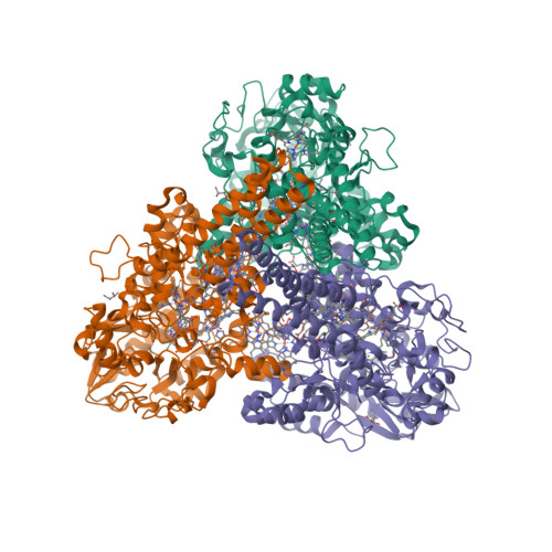
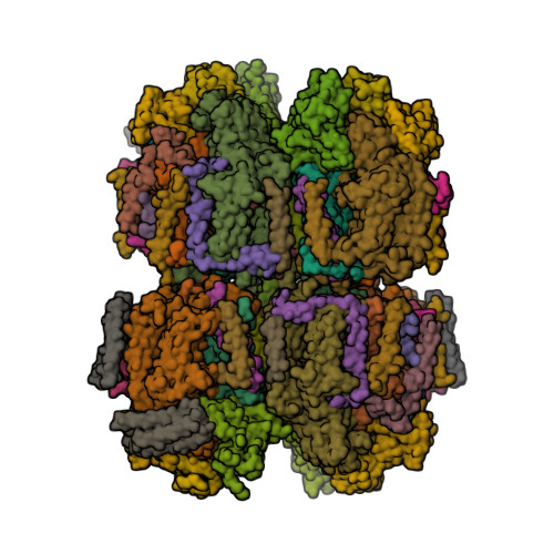
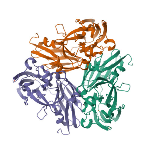
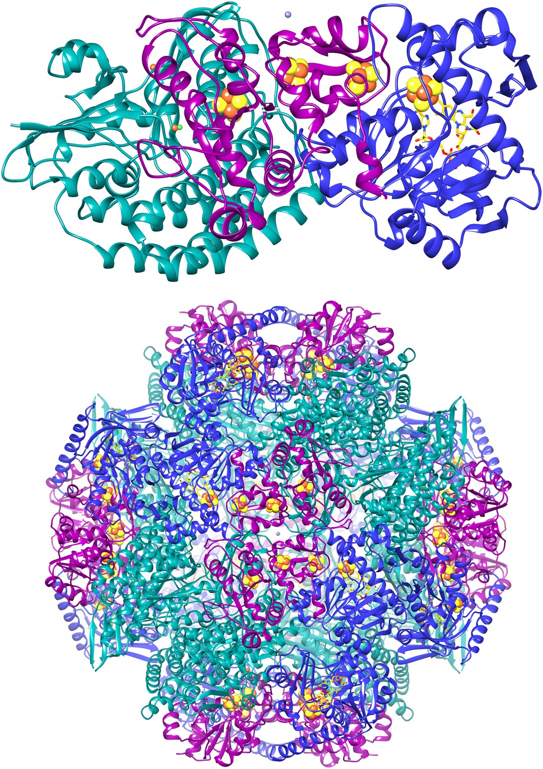
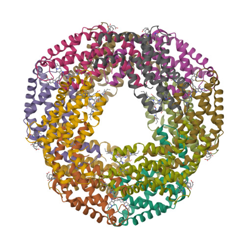
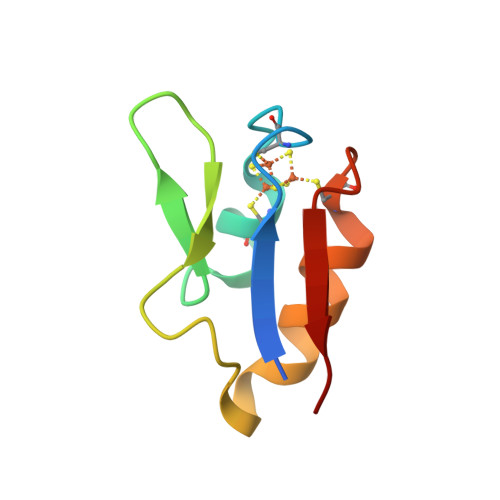
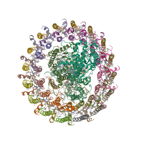
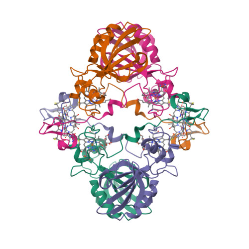
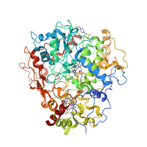
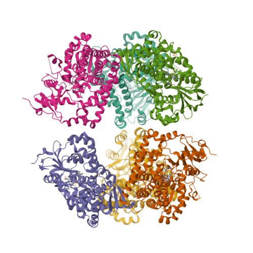
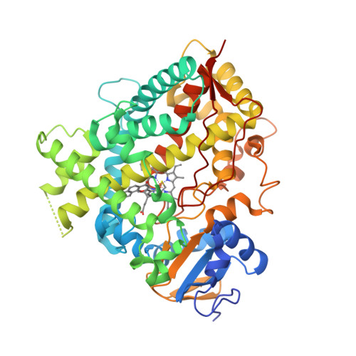
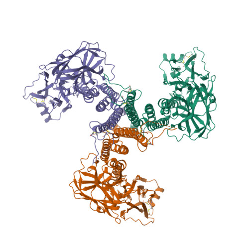
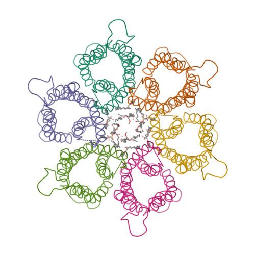
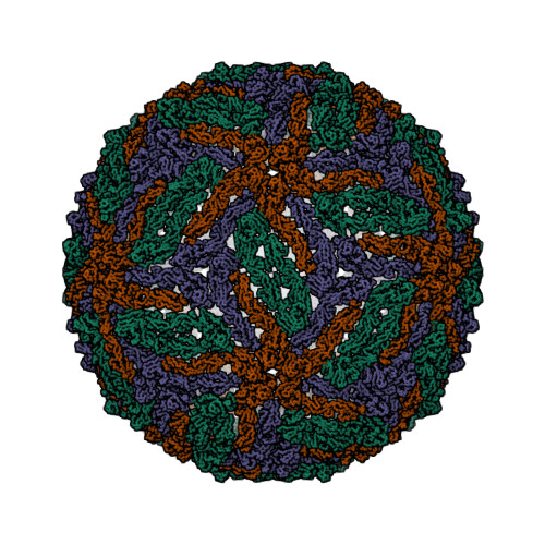
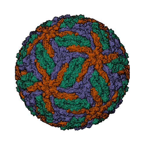
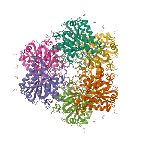
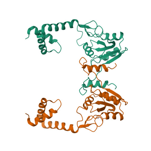
Wagner et al. [1] determined the X-ray structures of a Fwd enzyme from the thermophilic methanogenic archaeon Methanothermobacter wolfeii in several crystal forms [2—4]. To any bioinorganic chemist this metalloprotein should look like a treasure trove: every FwdABCDFG heterohexamer has got a mononuclear tungsten centre, a dinuclear zinc centre, and quite a few iron—sulphur clusters. The enzyme exists as either a dimer or a tetramer of the FwdABCDFG heterohexamers. The authors suggest that the 24-meric complex (FwdABCDFG)4 is a physiologically active form. It contains 46 (yes, forty-six) [Fe4S4] clusters which form an electron wire between the redox-active tungsten centres. The function of this wire remains unclear though.

  1. Wagner, T., Ermler, U. and Shima, S. (2016) The methanogenic CO2 reducing-and-fixing enzyme is bifunctional and contains 46 [4Fe-4S] clusters. Science 354, 114—117.
  2. PDB:5T5I
  3. PDB:5T5M
  4. PDB:5T61

Sunday, April 03, 2016

Octahaem sulphite reductase MccA

The epsilonproteobacterium Wolinella succinogenes is able to grow by sulphite respiration with formate as electron donor [1], thanks to the octahaem cytochrome c MccA that catalyses the six-electron reduction of sulphite to sulphide:

HSO3 + 6 H+ + 6 e → HS + 3 H2O

The crystal structure of MccA has been determined at 2.2 Å resolution [2, 3]. The enzyme exists as a homotrimer showing a novel fold and haem arrangement. The heterobimetallic active centre contains a Cu(I) ion and a haem c with a Fe—Cu distance of 4.4 Å [4].

a, W. succinogenes MccA binds its substrate sulfite in the dehydrated form, SO2, at the distal axial position of haem 2. At 3.2 Å distance from the sulphur atom, a Cu(I) ion is nearly linearly coordinated by residues C399 and C495.

b, In respiratory haem–copper oxidases, CuB is a redox-active species liganded by three histidine residues and juxtaposed to a haem a3 moiety. The arrangement, with a Fe–Cu distance of 4.9 Å, is optimized to bind O2 and peroxide in a bridging fashion (PDB:3ABM).

  1. Kern, M., Klotz, M.G. and Simon, J. (2011) The Wolinella succinogenes mcc gene cluster encodes an unconventional respiratory sulphite reduction system. Molecular Microbiology 82, 1515—1530.
  2. PDB:4RKM
  3. PDB:4RKN
  4. Hermann, B., Kern, M., La Pietra, L., Simon, J., Einsle, O. (2015) The octahaem MccA is a haem c-copper sulfite reductase. Nature 520, 706—709.

Saturday, February 27, 2016

First eukaryotic photosystem II solved at 2.76 Å

The water-splitting reaction of photosynthesis is catalysed by photosystem II (PSII), a large protein complex located in thylakoid membranes of organisms ranging from cyanobacteria to higher plants [1]. During the last 15 years, a number of crystal structures of PSII from cyanobacteria have been solved. However, no structures of PSII from eukaryots have been reported until now, partly due to the instability of eukaryotic PSII upon isolation. Ago et al. [2] solved the structure of PSII from a red alga Cyanidium caldarium at 2.76 Å resolution [3]. This PSII contains four extrinsic proteins, including the three subunits found in cyanobacterial PSII and the fourth subunit PsbQ' homologous to the PsbQ protein of green algae and higher plants. Furthermore, two novel trans-membrane helices were found in the algal PSII which are not present in cyanobacterial PSII.

  1. Shen, J.-R. (2015) The structure of photosystem II and the mechanism of water oxidation in photosynthesis. Annual Review of Plant Biology 66, 23—48.
  2. Ago, H., Adachi, H., Umena, Y., Tashiro, T., Kawakami, K., Kamiya, N., Tian, L., Han, G., Kuang, T., Liu, Z., Wang, F., Zou, H., Enami, I., Miyano, M. and Shen, J.-R. (2016) Novel features of eukaryotic photosystem II revealed by its crystal structure analysis from a red alga. J. Biol. Chem. 291, 5676—5687.
  3. PDB:4YUU

Saturday, January 30, 2016

Crystal structure of the DNAzyme 9DB1

The first ever crystal structure of a deoxyribozyme has been solved at 2.8 Å resolution [1—3]. The work by researchers from Max Planck Institute for Biophysical Chemistry (Göttingen, Germany) also sheds light on a difference in catalytic mechanism of ribozymes and deoxyribozymes [4]:

Ribozymes use RNA’s 2´-hydroxyl groups, which are absent in DNA, for structural interactions or directly for catalysis. The new structure shows why the lack of these groups doesn’t diminish the catalytic activity of DNAzymes. The missing hydroxyls make DNA’s sugar-phosphate backbone more flexible, allowing acrobatic conformations that compensate for the absent hydroxyls in DNAzymes.

  1. Ponce-Salvatierra, A., Wawrzyniak-Turek, K., Steuerwald, U., Höbartner, C. and Pena, V. (2016) Crystal structure of a DNA catalyst. Nature 529, 231—234.
  2. PDB:5CKK
  3. PDB:5CKI
  4. Borman, S. (2016) After two decades of trying, scientists report first crystal structure of a DNAzyme. Chemical & Engineering News 94, issue 2, p. 3.

Thursday, November 19, 2015

A magnetic protein biocompass?

A team of scientists from Peking University report a putative magnetic receptor (MagR) protein in Drosophila, CG8198 [1]. MagR binds an iron—sulphur cluster and interacts with photoreceptor cryptochrome (Cry) proteins to form a multimeric magnetosensing rod-like complex. Assemblies of these rods were observed orienting themselves in a weak magnetic field. Qin et al. speculate that these structures may function like compasses in living organisms, although the mechanism of magnetoreception in vivo remains a mystery [2].

A complete Cry/MagR magnetosensor protein complex structure model with 10 Crys helically binding to the rod-like MagR polymer consisting of 20 MagRs.
  1. Qin, S., Yin, H., Yang, C., Dou, Y., Liu, Z., Zhang, P., Yu, H., Huang, Y., Feng, J., Hao, J., Hao, J., Deng, L., Yan, X., Dong, X., Zhao, Z., Jiang, T., Wang, H.-W., Luo, S.-J. and Xie, C. (2016) A magnetic protein biocompass. Nature Materials 15, 217–226.
  2. Cyranoski, D. (2015) Long-sought biological compass discovered: Protein complex offers explanation for how animals sense Earth’s magnetic pull. Nature 527, 283–284.

Sunday, September 27, 2015

Nitrite binding modes in CuNIR

Fukuda and Inoue [1] have determined the crystal structure of the C135A mutant of thermostable copper nitrite reductase (CuNIR) from Geobacillus thermodenitrificans in complex with nitrite to 1.55 Å resolution. Interestingly, this high-temperature (320 K) structure [2] displays a near-bidentate binding mode of nitrite distinct from a monodentate mode in a cryogenic structure [3]:

To our knowledge, this is the first case in which the difference in substrate binding modes between cryogenic and high-temperature structures has been visualized by crystallography.

The copper site geometries are given in Table 1.

Table 1 (adapted from [4])

Cu—Ligand Distances (Å) 3X1N (320 K) 3WKP (100 K)
I. Type 1 Cu—residue distances
T1Cu—H95Nδ1 2.08 2.14
T1Cu—H143Nδ1 2.01 1.96
T1Cu—M148Sδ 2.13 2.07
II. Type 2 Cu—residue distances
T2Cu—H100Nε2 2.07 1.96
T2Cu—H134Nε2 2.00 1.95
T2Cu—H294Nε2 1.98 2.01
T2Cu—water 2.02 n/a
III. Type 2 Cu—nitrite distances
T2Cu—Oproximal 2.13 1.97
T2Cu—N 2.21 2.85
T2Cu—Odistal 2.52 3.41
  1. Fukuda, Y. and Inoue, T. (2015) High-temperature and high-resolution crystallography of thermostable copper nitrite reductase. Chemical Communications 51, 6532—6535.
  2. PDB:3X1N
  3. PDB:3WKP
  4. Fukuda, Y. and Inoue, T. (2015) High-temperature and high-resolution crystallography of thermostable copper nitrite reductase. Electronic Supplementary Material.

Tuesday, September 23, 2014

Pseudomonas fluorescens PhoX

Alkaline phosphatases (EC 3.1.3.1) occur widely in nature and are found in all three domains of life [1]. The Escherichia coli PhoA enzyme has been extensively studied whereas PhoX family of alkaline phosphatases are only minimally characterised and show no sequence similarity to other phosphotransfer enzymes. Yong et al. [2] determined high-resolution crystal structures for native PhoX from Pseudomonas fluorescens [3] and for its complexes with phosphate [4], a nonhydrolysable ATP analogue adenosine-5′-[β,γ-methylene]triphosphate (AMP-PCP) [5], and the putative transition-state mimic vanadate [6]. The active site contains two antiferromagnetically coupled ferric ions (Fe3+), three calcium ions (Ca2+), and an oxo group bridging one Ca2+ and two Fe3+ ions.

Cartoon representation of P. fluorescens PhoX crystal structure.
The PhoX active site containing bound phosphate [1, Fig. 2c].
A model for the catalytic mechanism of PhoX [1, Fig. 3d].
The transition state is indicated with the double dagger (‡) symbol.
  1. Millán, J.L. (2006) Alkaline Phosphatases: Structure, substrate specificity and functional relatedness to other members of a large superfamily of enzymes. Purinergic Signalling 2, 335–341.
  2. Yong, S.C., Roversi, P., Lillington, J., Rodriguez, F., Krehenbrink, M., Zeldin, O.B., Garman, E.F., Lea, S.M. and Berks, B.C. (2014) A complex iron-calcium cofactor catalyzing phosphotransfer chemistry. Science 345, 1170—1173.
  3. PDB:4A9V
  4. PDB:4ALF
  5. PDB:4AMF
  6. PDB:3ZWU

Thursday, July 31, 2014

F420-reducing [NiFe]-hydrogenase at 1.7 Å

The F420-reducing [NiFe]-hydrogenase (FrhABG; EC 1.12.98.1) catalyses the reversible redox reaction between coenzyme F420 and H2. FrhABG is a group 3 [NiFe]-hydrogenase with a dodecameric quaternary structure recently revealed by high-resolution cryo-electron microscopy [1]. Vitt et al. report the crystal structure of FrhABG from Methanothermobacter marburgensis at 1.7 Å resolution [2, 3] and compare it with the structures of group 1 [NiFe]-hydrogenases, the only previously structurally characterised group.

  1. Allegretti, M., Mills, D.J., McMullan, G., Kühlbrandt, W. and Vonck, J. (2014) Atomic model of the F420-reducing [NiFe] hydrogenase by electron cryo-microscopy using a direct electron detector. eLife 3, e01963.
  2. Vitt, S., Ma, K., Warkentin, E., Moll, J., Pierik, A.J., Shima, S. and Ermler, U. (2014) The F420-reducing [NiFe]-hydrogenase complex from Methanothermobacter marburgensis, the first X-ray structure of a group 3 family member. J. Mol. Biol. 426, 2813—2826.
  3. PDB:4OMF

Monday, June 30, 2014

Phycocyanin against Alzheimer’s?

Could a light-harvesting protein phycocyanin be used as a novel drug against Alzheimer’s disease (AD) [1, 2]?

In the present study, intact hexameric phycocyanin was isolated and crystallized from the cyanobacterium Leptolyngbya sp. N62DM, and the structure was solved to a resolution of 2.6 Å. Molecular docking studies show that the phycocyanin αβ-dimer interacts with the enzyme β-secretase, which catalyzes the proteolysis of the amyloid precursor protein to form plaques. The molecular docking studies suggest that the interaction between phycocyanin and β-secretase is energetically more favorable than previously reported inhibitor-β-secretase interactions. Transgenic Caenorhabditis elegans worms, with a genotype to serve as an AD-model, were significantly protected by phycocyanin. Therefore, the present study provides a novel structure-based molecular mechanism of phycocyanin-mediated therapy against AD.
  1. Singh, N.K., Hasan, S.S., Kumar, J., Raj, I., Pathan, A.A., Parmar, A., Shakil, S., Gourinath, S. and Madamwar D. (2014) Crystal structure and interaction of phycocyanin with β-secretase: A putative therapy for Alzheimer's disease. CNS Neurol. Disord. Drug Targets 13, 691—698.
  2. PDB:4L1E

Saturday, May 24, 2014

[Fe3S4] ferredoxin from Rhodopseudomonas palustris

The crystal structure of a novel [Fe3S4] ferredoxin associated with CYP194A4 from Rhodopseudomonas palustris has been solved at 2.15 Å resolution [1—3]. The ferredoxin, HaPuxC, contains an atypical CXXHXXC(X)nCP iron-sulphur cluster-binding motif. HaPuxC is the first P450 electron-transfer partner of this type to be structurally characterised.

  1. Zhang, T., Zhang, A., Bell, S.G., Wong, L.-L. and Zhou, W. (2014) The structure of a novel electron-transfer ferredoxin from Rhodopseudomonas palustris HaA2 which contains a histidine residue in its iron-sulfur cluster-binding motif. Acta Crystallographica D70, 1453—1464.
  2. PDB:4ID8
  3. PDB:4OV1

Thursday, May 01, 2014

Tetracalcium octachromium(3+) strontium octacarbonate hexadecahydroxide sulfate pentaicosahydrate

The Polar Bear peninsula in Western Australia is one of the many places on this planet I never heard before. The reason I mention it now is that a new mineral named putnisite was discovered there, and this mineral caused a bit of a stir recently, for being “completely unique and unrelated to anything”. In fact, if you Google “Polar Bear peninsula”, all you find is putnisite.

In 2007, specimens of an unknown mineral forming purple crystals (a) were collected at the Polar Bear peninsula while prospecting for nickel and gold. The specimens were eventually forwarded to Peter Elliott, a research associate with the South Australian Museum, for examination.

(a)

Elliott et al. [2] report the composition and crystal structure of this unique mineral, named in honour of mineralogists Christine and Andrew Putnis of the Institut für Mineralogie, Universtität Münster, Germany. The compositional name for putnisite I come up with is “tetracalcium octachromium(3+) strontium octacarbonate hexadecahydroxide sulfate pentaicosahydrate”. Curiously, Mindat and Mineralienatlas give the molecular formula containing only 23 molecules of water.

(b)

The crystal structure (b) was determined from single-crystal X-ray diffraction data. Cr(OH)4O2 octahedra (red) link by edge-sharing to form an eight-membered ring. At the centre of each ring lies a decacoordinated Sr2+ cation (purple). The rings are decorated by carbonate triangles (green), each of which links by corner-sharing to two Cr(OH)4O2 octahedra. Rings are linked by Ca(H2O)4O4 polyhedra (blue) to form a sheet parallel to the (100) plane. Adjacent sheets are joined along the [100] direction by corner-sharing sulfate tetrahedra (yellow).

  1. Mills, R. (2014) New mineral shows nature’s infinite variability. Phys.org.
  2. Elliott, P., Giester, G., Rowe, R. and Pring, A. (2014) Putnisite, SrCa4Cr3+8(CO3)8SO4(OH)16·25H2O, a new mineral from Western Australia: description and crystal structure. Mineralogical Magazine 78, 131—144.

Tuesday, April 22, 2014

Thermochromatium tepidum LH1—RC complex at 3.0 Å

The light-harvesting core antenna (LH1) and the reaction centre (RC) of purple photosynthetic bacteria form a supramolecular complex (LH1—RC) to use sunlight energy in a highly efficient manner. Niwa et al. [1—4] report the first near-atomic structure of a LH1—RC complex, namely that of a Ca2+-bound complex from Thermochromatium tepidum. The RC is surrounded by 16 heterodimers of the LH1 αβ-subunit that form a completely closed structure. The Ca2+ ions are located at the periplasmic side of LH1. Thirty-two bacteriochlorophyll a and sixteen spirilloxanthin molecules in the LH1 ring form an elliptical assembly.

  1. Niwa, S., Yu, L.-J., Takeda, K., Hirano, Y., Kawakami, T., Wang-Otomo, Z.-Y. and Miki, K. (2014) Structure of the LH1—RC complex from Thermochromatium tepidum at 3.0 Å. Nature 508, 228—232.
  2. PDB:3WMM
  3. PDB:4V8K

Saturday, January 18, 2014

Sodium chloride revisited

Everybody knows that the formula of sodium chloride is NaCl. Right? Right. But recently, the team of Artem Oganov at Stony Brook University have shown that there are other stable types of crystalline sodium chloride. They have predicted several thermodynamically stable compounds: Na3Cl, Na2Cl, Na3Cl2, NaCl3, and NaCl7. Moreover, by utilising high-pressure techniques, they synthesised cubic and orthorhombic NaCl3 and two-dimensional tetragonal Na3Cl [1, 2].

NaCl3 (space group Pm3n)
Na3Cl (space group P4/mmm)

“One of these materials — Na3Cl — has a fascinating structure”, says Oganov. “It is comprised of layers of NaCl and layers of pure sodium. The NaCl layers act as insulators; the pure sodium layers conduct electricity” [3].

  1. Zhang, W., Oganov, A.R., Goncharov, A.F., Zhu, Q., Boulfelfel, S.E., Lyakhov, A.O., Stavrou, E., Somayazulu, M., Prakapenka, V.B. and Konôpková, Z. (2013) Unexpected stable stoichiometries of sodium chlorides. Science 342, 1502—1505; arXiv:1310.7674v1
  2. Ibáñez Insa, J. (2013) Reformulating table salt under pressure. Science 342, 1459—1460.
  3. SBU team discovers new compounds that challenge the foundation of chemistry. Stony Brook University Newsroom, December 19, 2013.

Tuesday, November 26, 2013

Magnetochrome-containing iron oxidase MamP

Magnetotactic bacteria (MTB) are a diverse group of prokaryotes that have a singular ability to align with geomagnetic field lines. This ability is due to special organelles called magnetosomes. Magnetosomes are composed of single-magnetic-domain nanocrystals of magnetite [Fe(II)Fe(III)2O4] or greigite [Fe(II)Fe(III)2S4] embedded in biological membrane.

“Magnetochrome” is a name proposed in 2012 by Marina Siponen and co-authors for a cytochrome domain conserved within all known MTB and not found in any other species to date [1]. Recently, the crystal structure of the magnetosome-associated protein MamP has been solved at 1.8 Å resolution [2—4]. The minimal unit of MamP is a dimer. Each monomer consists of a PDZ domain fused to two magnetochrome domains. It was also shown in an in vitro mineralisation experiment that MamP functions as an iron oxidase mediating the iron(III) ferrihydrite production from iron(II) [2]:

4Fe2+ + 7H2O → 2Fe2O3·H2O + 12H+ + 4e
  1. Siponen, M.I., Adryanczyk, G., Ginet, N., Arnoux, P. and Pignol, D. (2012) Magnetochrome: a c-type cytochrome domain specific to magnetotatic bacteria. Biochemical Society Transactions 40, 1319—1323.
  2. Siponen, M.I., Legrand, P., Widdrat, M., Jones, S.R., Zhang, W.-J., Chang, M.C.Y., Faivre, D., Arnoux, P. and Pignol, D. (2013) Structural insight into magnetochrome-mediated magnetite biomineralization. Nature 502, 681—684.
  3. PDB:4JJ0
  4. PDB:4JJ3

Saturday, October 26, 2013

The first viral cytochrome b5

A unicellular green alga Ostreococcus tauri is the smallest (less than 1 μm in diameter) free-living eukaryote yet described. Viruses that can infect high-light and low-light adapted strains of O. tauri have been isolated and their genomes sequenced. Interestingly, low-light-strain infecting virus (OtV-2) differ from the high-light-strain infecting viruses by encoding a potential cytochrome b5 [1]. This protein was cloned, biochemically characterised and its three-dimensional structure resolved [2, 3].

The absorption spectra of oxidised and reduced recombinant OtV-2 haemoprotein are almost identical to those of purified human cytochrome b5.

Absorbance spectra of purified recombinant human cytochrome b5 and OtV-2_201.

It was also shown that the protein can substitute for yeast cytochrome b5 in the CYP51-mediated sterol 14α-demethylation. Structurally, the viral protein is similar to other known cytochromes b5 but lacks a hydrophobic C-terminal anchor. Thus, the first virally encoded cytochrome b5 is also the first cytosolic cytochrome b5 characterised. However, the physiological role of viral cytochrome b5 remains unknown.

  1. UniProt:E4WM77
  2. Reid, E.L., Weynberg, K.D., Love, J., Isupov, M.N., Littlechild, J.A., Wilson, W.H., Kelly, S.L., Lamb, D.C. and Allen, M.J. (2013) Functional and structural characterisation of a viral cytochrome b5. FEBS Letters, 587, 3633—3639.
  3. PDB:4B8N

A structural representation of the OtV-2 cytochrome b5 (OtV-2_201) protein shown as a ribbon diagram.

Wednesday, July 24, 2013

Crystal structure of latex oxygenase RoxA

To date, two types of enzymes that are responsible for primary attack of polyisoprene in rubber-degrading microorganisms have been identified [1]. One is the latex clearing protein (Lcp), first isolated from Streptomyces sp., which does not have any metal ions or cofactors [2]. The other is the rubber oxygenase RoxA of Xanthomonas sp., a dihaem c-type cytochrome that cleaves cis-1,4-polyisoprene, the main constituent of natural rubber, to 12-oxo-4,8-dimethyltrideca-4,8-diene-1-al [3, 4]. The crystal structure of RoxA, solved at 1.8 Å resolution, was released today [5].

Latex Oxygenase RoxA @ PDB
  1. Birke, J., Hambsch, N., Schmitt, G., Altenbuchner, J. and Jendrossek, D. (2012) Phe317 is essential for rubber oxygenase RoxA activity. Applied and Environmental Microbiology 78, 7876—7883.
  2. Rose, K., Tenberge, K.B. and Steinbüchel, A. (2005) Identification and characterization of genes from Streptomyces sp. strain K30 responsible for clear zone formation on natural rubber latex and poly(cis-1,4-isoprene) rubber degradation. Biomacromolecules 6, 180—188.
  3. Braaz, R., Fischer, P. and Jendrossek, D. (2004) Novel type of heme-dependent oxygenase catalyzes oxidative cleavage of rubber (poly-cis-1,4-isoprene). Applied and Environmental Microbiology 70, 7388—7395.
  4. Schmitt, G., Seiffert, G., Kroneck, P.M.H., Braaz, R. and Jendrossek, D. (2010) Spectroscopic properties of rubber oxygenase RoxA from Xanthomonas sp., a new type of dihaem dioxygenase. Microbiology 156, 2537—2548.
  5. PDB:4B2N

Saturday, June 22, 2013

S-Adenosyl-S-carboxymethyl-L-homocysteine

A novel cofactor is not something that is discovered every day, or even every year. So we are lucky this year. The crystal structure of a putative methyltransferase CmoA from Escherichia coli reveals the presence of [(3S)-3-amino-3-carboxypropyl]{[(2S,3S,4R,5R)-5-(6-aminopurin-9-yl)-3,4-dihydroxyoxolan-2-yl]methyl}(carboxymethyl)sulfanium, aka S-adenosyl-S-carboxymethyl-L-homocysteine, aka SCM-SAH [1—3]. Moreover, it was suggested that “a number of enzymes that have previously been annotated as SAM-dependent are in fact SCM-SAH-dependent” [1].

  1. Byrne, R.T., Whelan, F., Aller, P., Bird, L.E., Dowle, A., Lobley, C.M., Reddivari, Y., Nettleship, J.E., Owens, R.J., Antson, A.A. and Waterman, D.G. (2013) S-Adenosyl-S-carboxymethyl-L-homocysteine: a novel cofactor found in the putative tRNA-modifying enzyme CmoA. Acta Crystallographica D69, 1090—1098.
  2. PDB:4GEK
  3. PDB:4IWN

Wednesday, May 15, 2013

Aldosterone synthase structures

Earlier this year [1], the crystal structures of human aldosterone synthase (CYP11B2) were solved in complex with a substrate 11-deoxycorticosterone [2] and an inhibitor fadrozole [3].

  1. Strushkevich, N., Gilep, A.A., Shen, L., Arrowsmith, C.H., Edwards, A.M., Usanov, S.A. and Park, H.-W. (2013) Structural insights into aldosterone synthase substrate specificity and targeted inhibition. Molecular Endocrinology 27, 315—324.
  2. PDB:4DVQ
  3. PDB:4FDH

Thursday, February 28, 2013

The first CYP1A1 structure

CYP1A1 was one of the first P450 enzymes to be characterised and, as its name indicates, holds the first place in the systematic nomenclature of P450s [1]. However, it was not until last year that the first crystal structure of human CYP1A1 in complex with α-naphthoflavone has been determined at 2.6 Å resolution [2]. The structure [3] is released this week.

  1. Nebert, D.W., Nelson, D.R., Coon, M.J., Estabrook, R.W., Feyereisen, R., Fujii-Kuriyama, Y., Gonzalez, F.J., Guengerich, F.P., Gunsalus, I.C., Johnson, E.F., Loper, J.C., Sato, R., Waterman, M.R. and Waxman, D.J. (1991) The P450 superfamily: update on new sequences, gene mapping, and recommended nomenclature. DNA Cell Biol. 10, 1—14.
  2. Walsh, A.A., Szklarz, G.D. and Scott, E.E. (2012) Cytochrome P450 1A1 structure and utility in predicting drug and xenobiotic metabolism. Proceedings of the 19th International Symposium on Microsomes and Drug Oxidations and 12th European ISSX Meeting, Noordwijk, The Netherlands, 17—21 June 2012.
  3. PDB:4I8V

Monday, December 24, 2012

Macromolecular Xmas decorations

This year I don’t have a Christmas tree. (There aren’t many in Fuerteventura.) But I’ve got some models from this week’s new PDB structures [1—5] which look Christmassy enough to decorate my blog. Merry Christmas and a happy New Year everybody!


  1. Hui, F., Scheib, U., Hu, Y., Sommer, R.J., Aroian, R.V. and Ghosh, P. (2012) Structure and glycolipid binding properties of the nematicidal protein Cry5B. Biochemistry 51, 9911—9921.
  2. Lence, E., Tizón, L., Otero, J.M., Peón, A., Prazeres, V.F.V., Llamas-Saiz, A.L., Fox, G.C., van Raaij, M.J., Lamb, H., Hawkins, A.R. and González-Bello, C. (2013) Mechanistic basis of the inhibition of type II dehydroquinase by (2S)- and (2R)-2-benzyl-3-dehydroquinic acids. ACS Chem. Biol. 8, 568—577.
  3. Strugatsky, D., McNulty, R., Munson, K., Chen, C.-K., Soltis, S.M., Sachs, G. and Luecke, H. (2013) Structure of the proton-gated urea channel from the gastric pathogen Helicobacter pylori. Nature 493, 255—258.
  4. Tang, Q., Gao, P., Liu, Y.-P., Gao, A., An, X.-M., Liu, S., Yan, X.-X. and Liang, D.-C. (2012) RecOR complex including RecR N-N dimer and RecO monomer displays a high affinity for ssDNA. Nucleic Acids Res. 40, 11115—11125.
  5. Ziervogel, B.K. and Roux, B. (2013) The binding of antibiotics in OmpF porin. Structure 21, 76—87.