Showing posts with label enzymes. Show all posts

Sunday, April 03, 2016

Octahaem sulphite reductase MccA

The epsilonproteobacterium Wolinella succinogenes is able to grow by sulphite respiration with formate as electron donor [1], thanks to the octahaem cytochrome c MccA that catalyses the six-electron reduction of sulphite to sulphide:

HSO3 + 6 H+ + 6 e → HS + 3 H2O

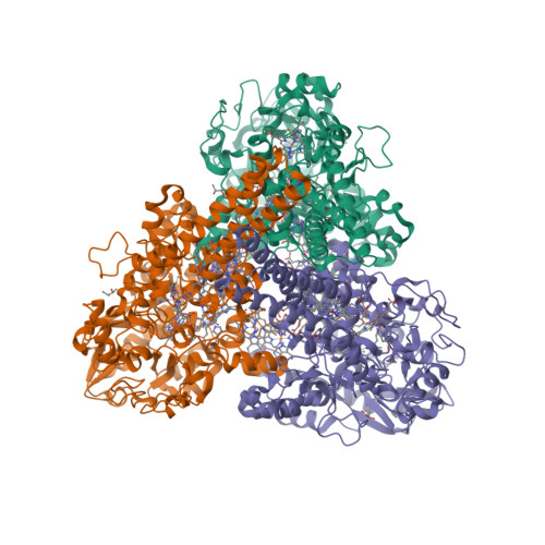
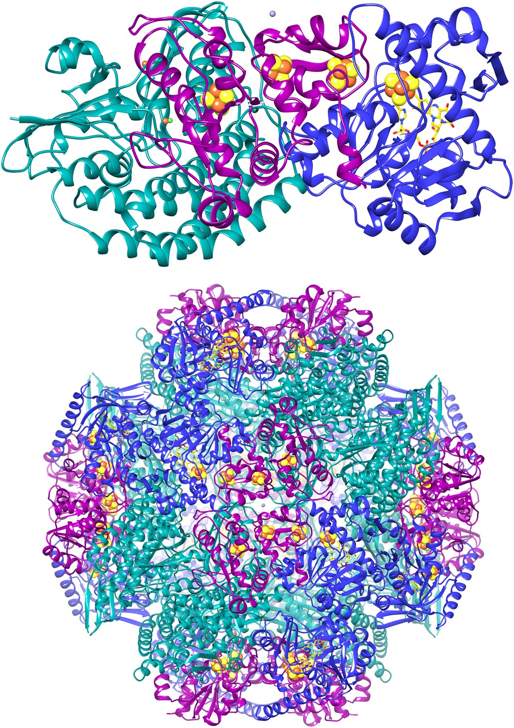
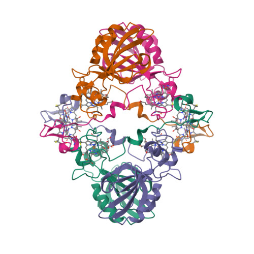
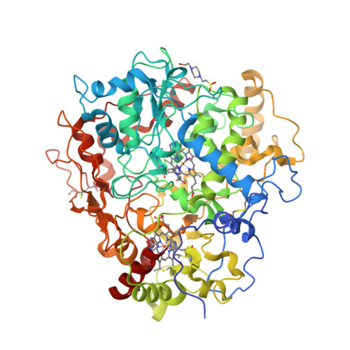
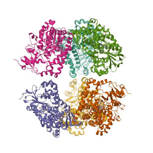
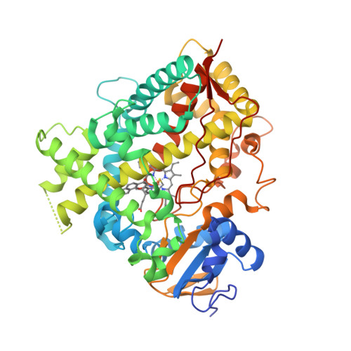
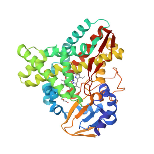
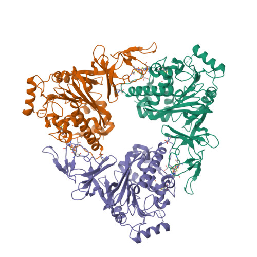
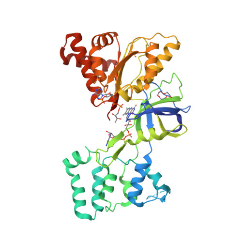
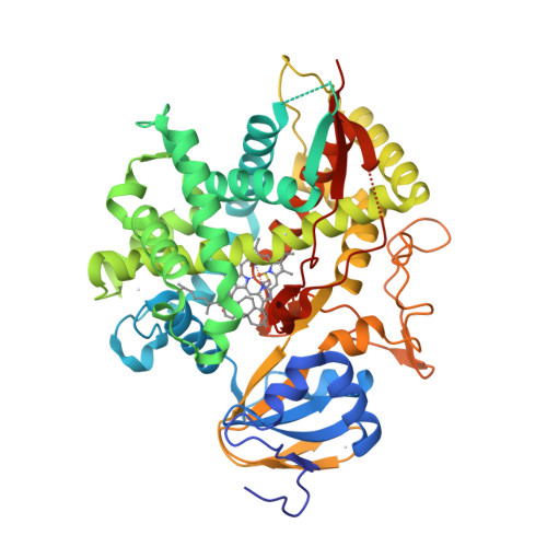
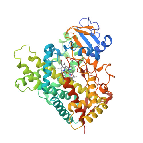
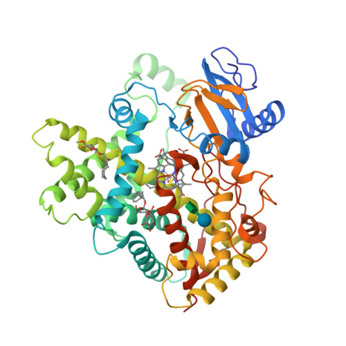
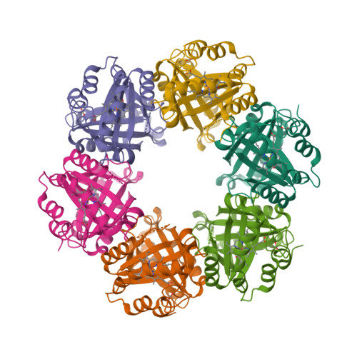
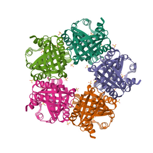
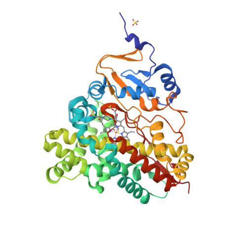
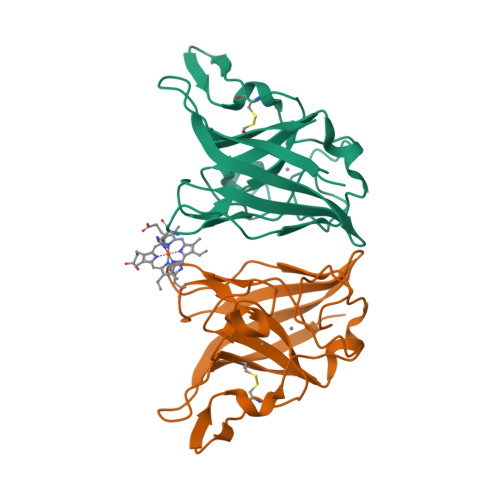
The crystal structure of MccA has been determined at 2.2 Å resolution [2, 3]. The enzyme exists as a homotrimer showing a novel fold and haem arrangement. The heterobimetallic active centre contains a Cu(I) ion and a haem c with a Fe—Cu distance of 4.4 Å [4].

a, W. succinogenes MccA binds its substrate sulfite in the dehydrated form, SO2, at the distal axial position of haem 2. At 3.2 Å distance from the sulphur atom, a Cu(I) ion is nearly linearly coordinated by residues C399 and C495.

b, In respiratory haem–copper oxidases, CuB is a redox-active species liganded by three histidine residues and juxtaposed to a haem a3 moiety. The arrangement, with a Fe–Cu distance of 4.9 Å, is optimized to bind O2 and peroxide in a bridging fashion (PDB:3ABM).

  1. Kern, M., Klotz, M.G. and Simon, J. (2011) The Wolinella succinogenes mcc gene cluster encodes an unconventional respiratory sulphite reduction system. Molecular Microbiology 82, 1515—1530.
  2. PDB:4RKM
  3. PDB:4RKN
  4. Hermann, B., Kern, M., La Pietra, L., Simon, J., Einsle, O. (2015) The octahaem MccA is a haem c-copper sulfite reductase. Nature 520, 706—709.

Tuesday, September 23, 2014

Pseudomonas fluorescens PhoX

Alkaline phosphatases (EC 3.1.3.1) occur widely in nature and are found in all three domains of life [1]. The Escherichia coli PhoA enzyme has been extensively studied whereas PhoX family of alkaline phosphatases are only minimally characterised and show no sequence similarity to other phosphotransfer enzymes. Yong et al. [2] determined high-resolution crystal structures for native PhoX from Pseudomonas fluorescens [3] and for its complexes with phosphate [4], a nonhydrolysable ATP analogue adenosine-5′-[β,γ-methylene]triphosphate (AMP-PCP) [5], and the putative transition-state mimic vanadate [6]. The active site contains two antiferromagnetically coupled ferric ions (Fe3+), three calcium ions (Ca2+), and an oxo group bridging one Ca2+ and two Fe3+ ions.

Cartoon representation of P. fluorescens PhoX crystal structure.
The PhoX active site containing bound phosphate [1, Fig. 2c].
A model for the catalytic mechanism of PhoX [1, Fig. 3d].
The transition state is indicated with the double dagger (‡) symbol.
  1. Millán, J.L. (2006) Alkaline Phosphatases: Structure, substrate specificity and functional relatedness to other members of a large superfamily of enzymes. Purinergic Signalling 2, 335–341.
  2. Yong, S.C., Roversi, P., Lillington, J., Rodriguez, F., Krehenbrink, M., Zeldin, O.B., Garman, E.F., Lea, S.M. and Berks, B.C. (2014) A complex iron-calcium cofactor catalyzing phosphotransfer chemistry. Science 345, 1170—1173.
  3. PDB:4A9V
  4. PDB:4ALF
  5. PDB:4AMF
  6. PDB:3ZWU

Thursday, July 31, 2014

F420-reducing [NiFe]-hydrogenase at 1.7 Å

The F420-reducing [NiFe]-hydrogenase (FrhABG; EC 1.12.98.1) catalyses the reversible redox reaction between coenzyme F420 and H2. FrhABG is a group 3 [NiFe]-hydrogenase with a dodecameric quaternary structure recently revealed by high-resolution cryo-electron microscopy [1]. Vitt et al. report the crystal structure of FrhABG from Methanothermobacter marburgensis at 1.7 Å resolution [2, 3] and compare it with the structures of group 1 [NiFe]-hydrogenases, the only previously structurally characterised group.

  1. Allegretti, M., Mills, D.J., McMullan, G., Kühlbrandt, W. and Vonck, J. (2014) Atomic model of the F420-reducing [NiFe] hydrogenase by electron cryo-microscopy using a direct electron detector. eLife 3, e01963.
  2. Vitt, S., Ma, K., Warkentin, E., Moll, J., Pierik, A.J., Shima, S. and Ermler, U. (2014) The F420-reducing [NiFe]-hydrogenase complex from Methanothermobacter marburgensis, the first X-ray structure of a group 3 family member. J. Mol. Biol. 426, 2813—2826.
  3. PDB:4OMF

Tuesday, November 26, 2013

Magnetochrome-containing iron oxidase MamP

Magnetotactic bacteria (MTB) are a diverse group of prokaryotes that have a singular ability to align with geomagnetic field lines. This ability is due to special organelles called magnetosomes. Magnetosomes are composed of single-magnetic-domain nanocrystals of magnetite [Fe(II)Fe(III)2O4] or greigite [Fe(II)Fe(III)2S4] embedded in biological membrane.

“Magnetochrome” is a name proposed in 2012 by Marina Siponen and co-authors for a cytochrome domain conserved within all known MTB and not found in any other species to date [1]. Recently, the crystal structure of the magnetosome-associated protein MamP has been solved at 1.8 Å resolution [2—4]. The minimal unit of MamP is a dimer. Each monomer consists of a PDZ domain fused to two magnetochrome domains. It was also shown in an in vitro mineralisation experiment that MamP functions as an iron oxidase mediating the iron(III) ferrihydrite production from iron(II) [2]:

4Fe2+ + 7H2O → 2Fe2O3·H2O + 12H+ + 4e
  1. Siponen, M.I., Adryanczyk, G., Ginet, N., Arnoux, P. and Pignol, D. (2012) Magnetochrome: a c-type cytochrome domain specific to magnetotatic bacteria. Biochemical Society Transactions 40, 1319—1323.
  2. Siponen, M.I., Legrand, P., Widdrat, M., Jones, S.R., Zhang, W.-J., Chang, M.C.Y., Faivre, D., Arnoux, P. and Pignol, D. (2013) Structural insight into magnetochrome-mediated magnetite biomineralization. Nature 502, 681—684.
  3. PDB:4JJ0
  4. PDB:4JJ3

Wednesday, July 24, 2013

Crystal structure of latex oxygenase RoxA

To date, two types of enzymes that are responsible for primary attack of polyisoprene in rubber-degrading microorganisms have been identified [1]. One is the latex clearing protein (Lcp), first isolated from Streptomyces sp., which does not have any metal ions or cofactors [2]. The other is the rubber oxygenase RoxA of Xanthomonas sp., a dihaem c-type cytochrome that cleaves cis-1,4-polyisoprene, the main constituent of natural rubber, to 12-oxo-4,8-dimethyltrideca-4,8-diene-1-al [3, 4]. The crystal structure of RoxA, solved at 1.8 Å resolution, was released today [5].

Latex Oxygenase RoxA @ PDB
  1. Birke, J., Hambsch, N., Schmitt, G., Altenbuchner, J. and Jendrossek, D. (2012) Phe317 is essential for rubber oxygenase RoxA activity. Applied and Environmental Microbiology 78, 7876—7883.
  2. Rose, K., Tenberge, K.B. and Steinbüchel, A. (2005) Identification and characterization of genes from Streptomyces sp. strain K30 responsible for clear zone formation on natural rubber latex and poly(cis-1,4-isoprene) rubber degradation. Biomacromolecules 6, 180—188.
  3. Braaz, R., Fischer, P. and Jendrossek, D. (2004) Novel type of heme-dependent oxygenase catalyzes oxidative cleavage of rubber (poly-cis-1,4-isoprene). Applied and Environmental Microbiology 70, 7388—7395.
  4. Schmitt, G., Seiffert, G., Kroneck, P.M.H., Braaz, R. and Jendrossek, D. (2010) Spectroscopic properties of rubber oxygenase RoxA from Xanthomonas sp., a new type of dihaem dioxygenase. Microbiology 156, 2537—2548.
  5. PDB:4B2N

Wednesday, May 15, 2013

Aldosterone synthase structures

Earlier this year [1], the crystal structures of human aldosterone synthase (CYP11B2) were solved in complex with a substrate 11-deoxycorticosterone [2] and an inhibitor fadrozole [3].

  1. Strushkevich, N., Gilep, A.A., Shen, L., Arrowsmith, C.H., Edwards, A.M., Usanov, S.A. and Park, H.-W. (2013) Structural insights into aldosterone synthase substrate specificity and targeted inhibition. Molecular Endocrinology 27, 315—324.
  2. PDB:4DVQ
  3. PDB:4FDH

Thursday, February 28, 2013

The first CYP1A1 structure

CYP1A1 was one of the first P450 enzymes to be characterised and, as its name indicates, holds the first place in the systematic nomenclature of P450s [1]. However, it was not until last year that the first crystal structure of human CYP1A1 in complex with α-naphthoflavone has been determined at 2.6 Å resolution [2]. The structure [3] is released this week.

  1. Nebert, D.W., Nelson, D.R., Coon, M.J., Estabrook, R.W., Feyereisen, R., Fujii-Kuriyama, Y., Gonzalez, F.J., Guengerich, F.P., Gunsalus, I.C., Johnson, E.F., Loper, J.C., Sato, R., Waterman, M.R. and Waxman, D.J. (1991) The P450 superfamily: update on new sequences, gene mapping, and recommended nomenclature. DNA Cell Biol. 10, 1—14.
  2. Walsh, A.A., Szklarz, G.D. and Scott, E.E. (2012) Cytochrome P450 1A1 structure and utility in predicting drug and xenobiotic metabolism. Proceedings of the 19th International Symposium on Microsomes and Drug Oxidations and 12th European ISSX Meeting, Noordwijk, The Netherlands, 17—21 June 2012.
  3. PDB:4I8V

Thursday, May 10, 2012

P450-flavodoxin fusion enzyme XplA

XplA is a P450-flavodoxin fusion enzyme that mediates the metabolism of the military explosive RDX (1,3,5-trinitro-1,3,5-triazinane) in Rhodococcus rhodochrous 11Y [1]. Bui et al. have conducted a detailed spectroscopic and crystallographic study of this unusual hemoflavoprotein [2, 3].

The XplA P450 has evolved as a reductase (rather than oxidase) of RDX and structural alterations to its heme- and FMN-binding domains have led to reduction potentials for low-spin heme iron Fe3+/Fe2+ and FMNSQ/HQ couples being much more positive than those seen in typical P450s and flavodoxins, but consistent with non-oxidative P450 catalysis. These evolutionary steps have also led to a constricted P450 active site with high affinity for RDX (but also for the small heterocyclic inhibitor imidazole), and also to substantially diminished affinity for FMN in the flavodoxin domain.

  1. Rylott, E.L., Jackson, R.G., Sabbadin, F., Seth-Smith, H.M.B., Edwards, J., Chong, C.S., Strand, S.E., Grogan, G. and Bruce, N.C. (2011) The explosive-degrading cytochrome P450 XplA: biochemistry, structural features and prospects for bioremediation. Biochim. Biophys. Acta 1814, 230—236.
  2. Bui, S.H., McLean, K.J., Cheesman, M.R., Bradley, J.M., Rigby, S.E.J., Levy, C.W., Leys, D. and Munro, A.W. (2012) Unusual spectroscopic and ligand binding properties of the cytochrome P450-flavodoxin fusion enzyme XplA. J. Biol. Chem. 287, 19699—19714.
  3. PDB:4EP6

Tuesday, April 24, 2012

Stachydrine demethylase

Crystal structures were determined for the Rieske-type monooxygenase, stachydrine demethylase, in the unliganded state (at 1.6 Å) and in the product complex (at 2.2 Å) [1—3].

  1. Daughtry, K.D., Xiao, Y., Stoner-Ma, D., Cho, E., Orville, A.M., Liu, P. and Allen, K.N. (2012) Quaternary ammonium oxidative demethylation: X-ray crystallographic, resonance Raman, and UV-visible spectroscopic analysis of a Rieske-type demethylase. J. Am. Chem. Soc. 134, 2823—2834.
  2. PDB:3VCA
  3. PDB:3VCP

Sunday, March 25, 2012

FAD/NADPH-domain of flavocytochrome P450 BM3

The crystal structure of the FAD/NADPH-binding domain of the Bacillus megaterium flavocytochrome P450 BM3 has been solved in both the absence and presence of the ligand NADP+ [1—3].

  1. Joyce, M.G., Ekanem, I.S., Roitel, O., Dunford, A.J., Neeli, R., Girvan, H.M., Baker, G.J., Curtis, R.A., Munro, A.W. and Leys, D. (2012) The crystal structure of the FAD/NADPH-binding domain of flavocytochrome P450 BM3. FEBS J. 279, 1694—1706.
  2. PDB:4DQK
  3. PDB:4DQL

Friday, July 29, 2011

Saturday, June 18, 2011

Open and closed P450 2B4

The crystal structures of rabbit P450 2B4 covalently bound to the mechanism-based inactivator 4-tert-butylphenylacetylene in closed (a) and open (b) conformations have been solved [1].


(a)

(b)

  1. Gay, S.C., Zhang, H., Wilderman, P.R., Roberts, A.G., Liu, T., Li, S., Lin, H.-l., Zhang, Q., Woods, V.L., Jr., Stout, C.D., Hollenberg, P.F. and Halpert, J.R. (2011) Structural analysis of mammalian cytochrome P450 2B4 covalently bound to the mechanism-based inactivator tert-butylphenylacetylene: insight into partial enzymatic activity. Biochemistry 50, 4903—4911.

Tuesday, March 22, 2011

Chlorite dismutase

Photosynthesis is not the only dioxygen-evolving biological process. For instance, chlorite dismutase (Cld; EC 1.13.11.49) catalyses the production of O2 from chlorite (1):

ClO2 → Cl + O2(1)

The reaction (1) is not really disproportionation, and NC-IUBMB made a valid point that the term “chlorite dismutase” is “misleading”. Even so, the NC-IUBMB-approved, sorry, “accepted” name “chlorite O2-lyase” for an oxidoreductase is equally absurd; I am going to ignore it.

Chlorite dismutase from Azospira oryzae exists as a homohexamer [1] while Cld from Dechloromonas aromatica [2] and enzyme from Candidatus Nitrospira defluvii [3] are homopentamers.

The active site contains a single haem group [Fe(ppIX)] coordinated by a proximal histidine residue. Goblirsch et al. [2] propose the mechanism where the reaction of chlorite within the distal pocket of Cld generates hypochlorite (ClO) and a compound I intermediate [Fe(ppIX)O] (1a). Then ClO rebounds with compound I forming the chloride and dioxygen (1b):

ClO2 + [Fe(ppIX)] → ClO + [Fe(ppIX)O](1a)
ClO + [Fe(ppIX)O] → Cl + O2 + [Fe(ppIX)](1b)
  1. de Geus, D.C., Thomassen, E.A.J., Hagedoorn, P.-L., Pannu, N.S., van Duijn, E. and Abrahams, J.P. (2009) Crystal structure of chlorite dismutase, a detoxifying enzyme producing molecular oxygen. J. Mol. Biol. 387, 192—206.
  2. Goblirsch, B.R., Streit, B.R., DuBois, J.L. and Wilmot, C.W. (2010) Structural features promoting dioxygen production by Dechloromonas aromatica chlorite dismutase. J. Biol. Inorg. Chem. 15, 879—888.
  3. Kostan, J., Sjöblom, B., Maixner, F., Mlynek, G., Furtmüller, P.G., Obinger, C., Wagner, M., Daims, H. and Djinović-Carugo, K. (2010) Structural and functional characterisation of the chlorite dismutase from the nitrite-oxidizing bacterium “Candidatus Nitrospira defluvii”: Identification of a catalytically important amino acid residue. J. Struct. Biol. 172, 331—342.

Thursday, March 17, 2011

P450 monooxygenase AurH from S. thioluteus

The first crystal structures of the unique P450 monooxygenase AurH from Streptomyces thioluteus have been solved [1].

  1. Zocher, G., Richter, M.E.A., Mueller, U. and Hertweck, C. (2011) Structural fine-tuning of a multifunctional cytochrome P450 monooxygenase. J. Am. Chem. Soc. 133, 2292—2302.

Tuesday, August 18, 2009

Deferrochelatase

I always thought that ferrochelatase (EC 4.99.1.1) has an absurd systematic name: “protoheme ferro-lyase (protoporphyrin-forming)”. Why? According to Enzyme Nomenclature,

Lyases are enzymes cleaving C—C, C—O, C—N and other bonds <in other words, any bond> by other means than by hydrolysis or oxidation.

“Protoheme ferro-lyase” implies that the reaction goes in the direction:

protoheme + 2 H+ → protoporphyrin + Fe2+(a)

while ferrochelatase, in fact, catalyses the reverse reaction:

protoporphyrin + Fe2+ → protoheme + 2 H+(b)

Usually, to release iron from protoheme, you have to break it. Heme oxygenase (EC 11.14.14.18) does it by sequential oxidation of heme into the linear tetrapyrrole, biliverdin.

However, this paper demonstrates that there could be another way to do it.

Until today, all known enzymes performing iron extraction from heme did so through the rupture of the tetrapyrrol skeleton. Here, we identified 2 Escherichia coli paralogs, YfeX and EfeB, without any previously known physiological functions. YfeX and EfeB promote iron extraction from heme preserving the tetrapyrrol ring intact. This novel enzymatic reaction corresponds to the deferrochelation of the heme. YfeX and EfeB are the sole proteins able to provide iron from exogenous heme sources to E. coli.

Thus, deferrochelatase catalyses the reaction (a) and, indeed, can be named “protoheme ferro-lyase (protoporphyrin-forming)”.

Sunday, February 22, 2009

Copper, zinc and haem in superoxide dismutase

Why bacterioferritin needs a haem anyway? It’s a very good question, and so far it does not have any good answer. The shortest one is, we don’t really know. There are ferritins (in Escherichia coli as well) having exactly the same architecture which do not bind any haem. Bacterioferritin is not alone: Cu,Zn superoxide dismutase from Haemophilus ducreyi contains not only, as one may guess, copper and zinc (all Cu,Zn-SODs do), but also, for no reason, a haem. Once again, it is bound at the dimer interface, although this time it is asymmetrically bound with two different histidine residues provided by the two subunits. The authors show that the introduction of only three mutations at the dimer interface of the Cu,Zn-SOD from a related species, Haemophilus parainfluenzae, is sufficient to induce haem-binding ability. However, it does not change anything: we still don’t know why the haem is there.