Showing posts with label bioinorganic chemistry. Show all posts

Thursday, September 10, 2026

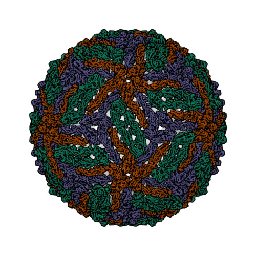
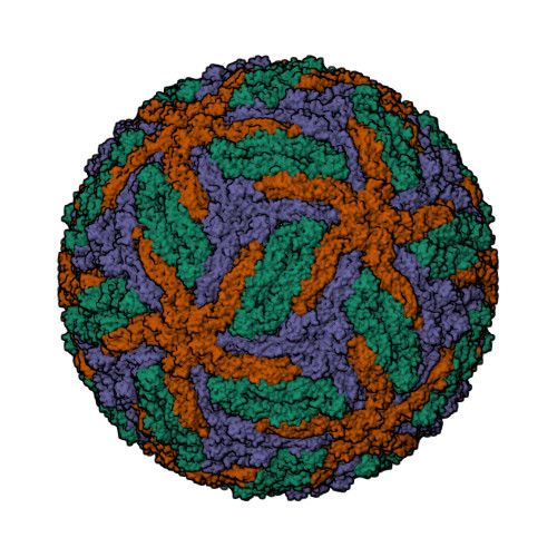
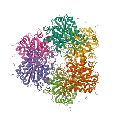
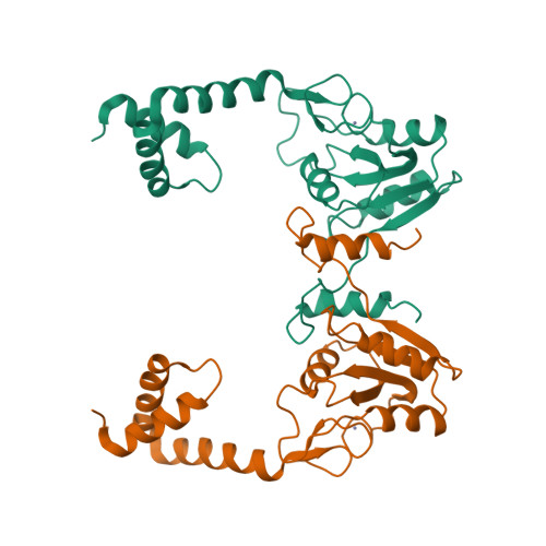
Pyrococcus abyssi rubredoxin at 0.43 Å

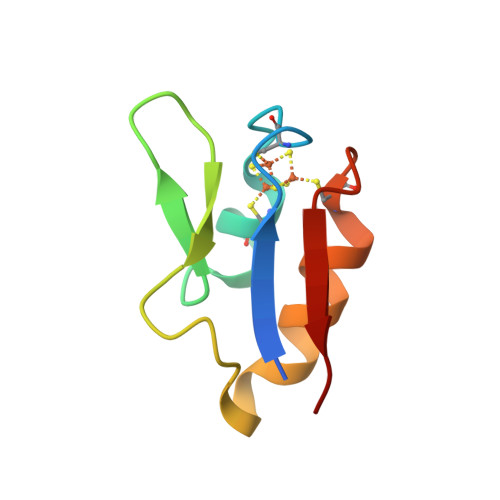
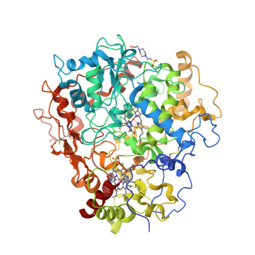
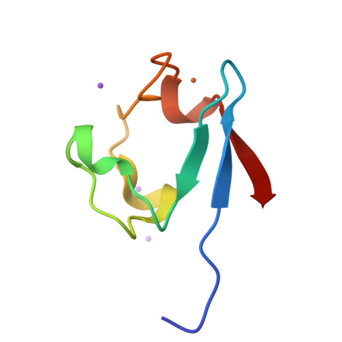
Paknia et al. report the X-ray crystal structure of rubredoxin from the archaeon Pyrococcus abyssi rubredoxin solved at a resolution of 0.43 Å [1, 2]. According to the authors, it “represents the highest resolution protein structure yet determined”. Currently, the PDB contains only 20 entries, apart from the reported ones [3, 4], at a resolution better than 0.7 Å.

  1. Paknia, E., Flensburg, C., Chodkiewicz, M.L., Fogh, R.H., Keller, P., Vonrhein, C., Schulze-Briese, C., Dominiak, P.M., Bourenkov, G., Bricogne, G. and Chari, A. (2026) Towards routine accurate electron-density studies of biological macromolecules. Acta Crystallographica D82, 1044—1055.
  2. Kuhn, C. (2026) Every electron counts: most detailed protein structure to date revealed. Max Planck Institute for Multidisciplinary Sciences, Research News, 2 September 2026.
  3. PDB:30OR
  4. PDB:30OH

Tuesday, December 06, 2016

An electron wire in formylmethanofuran dehydrogenase

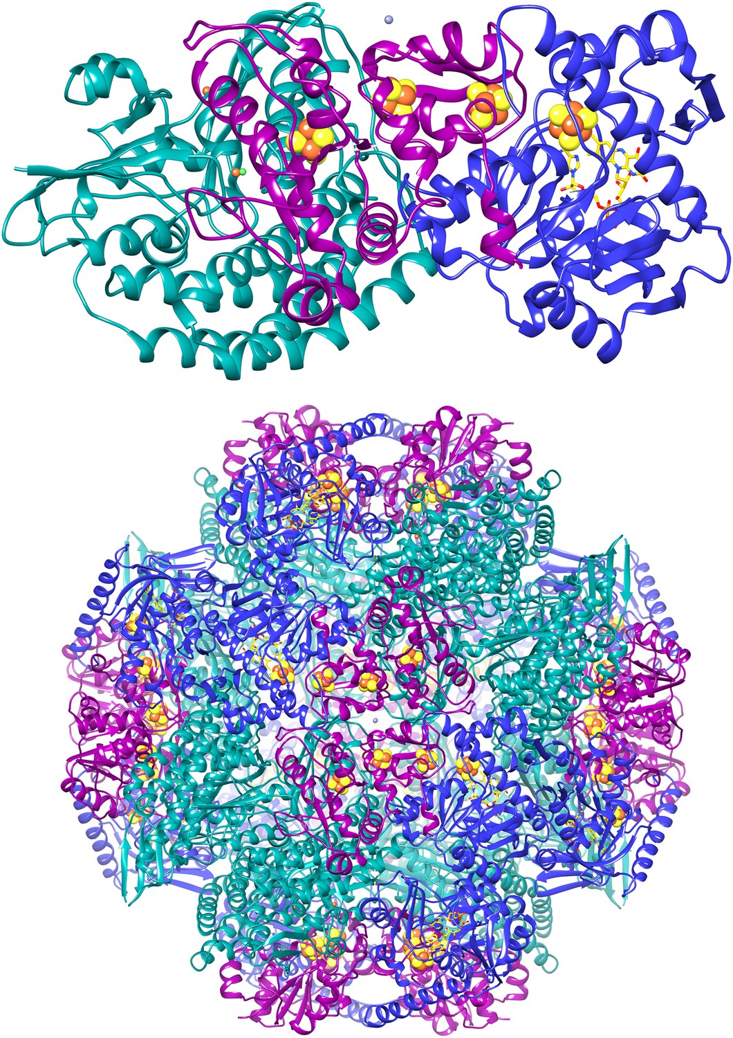
The first step of biological methane formation from carbon dioxide is the reduction of CO2 to form N-formylmethanofuran from methanofuran. This reaction is catalysed by formylmethanofuran dehydrogenase (EC 1.2.99.5). There are two types of this enzyme in methanogenic archaea, a tungsten iron—sulphur protein (Fwd) and a molybdenum iron—sulphur protein (Fmd).

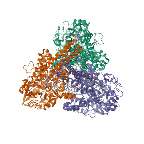
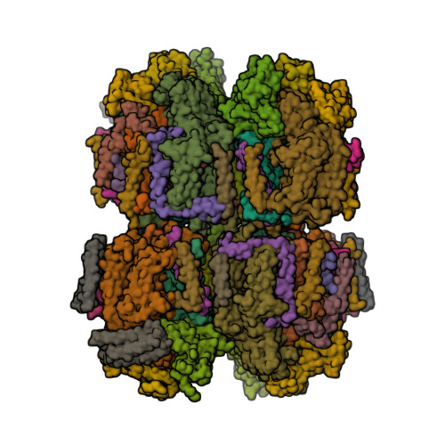
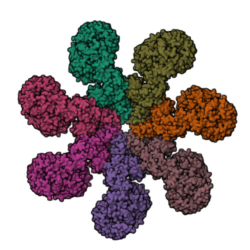
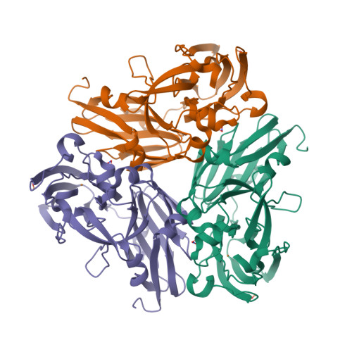
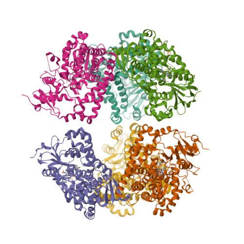
Wagner et al. [1] determined the X-ray structures of a Fwd enzyme from the thermophilic methanogenic archaeon Methanothermobacter wolfeii in several crystal forms [2—4]. To any bioinorganic chemist this metalloprotein should look like a treasure trove: every FwdABCDFG heterohexamer has got a mononuclear tungsten centre, a dinuclear zinc centre, and quite a few iron—sulphur clusters. The enzyme exists as either a dimer or a tetramer of the FwdABCDFG heterohexamers. The authors suggest that the 24-meric complex (FwdABCDFG)4 is a physiologically active form. It contains 46 (yes, forty-six) [Fe4S4] clusters which form an electron wire between the redox-active tungsten centres. The function of this wire remains unclear though.

  1. Wagner, T., Ermler, U. and Shima, S. (2016) The methanogenic CO2 reducing-and-fixing enzyme is bifunctional and contains 46 [4Fe-4S] clusters. Science 354, 114—117.
  2. PDB:5T5I
  3. PDB:5T5M
  4. PDB:5T61

Sunday, April 03, 2016

Octahaem sulphite reductase MccA

The epsilonproteobacterium Wolinella succinogenes is able to grow by sulphite respiration with formate as electron donor [1], thanks to the octahaem cytochrome c MccA that catalyses the six-electron reduction of sulphite to sulphide:

HSO3 + 6 H+ + 6 e → HS + 3 H2O

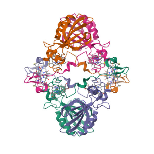
The crystal structure of MccA has been determined at 2.2 Å resolution [2, 3]. The enzyme exists as a homotrimer showing a novel fold and haem arrangement. The heterobimetallic active centre contains a Cu(I) ion and a haem c with a Fe—Cu distance of 4.4 Å [4].

a, W. succinogenes MccA binds its substrate sulfite in the dehydrated form, SO2, at the distal axial position of haem 2. At 3.2 Å distance from the sulphur atom, a Cu(I) ion is nearly linearly coordinated by residues C399 and C495.

b, In respiratory haem–copper oxidases, CuB is a redox-active species liganded by three histidine residues and juxtaposed to a haem a3 moiety. The arrangement, with a Fe–Cu distance of 4.9 Å, is optimized to bind O2 and peroxide in a bridging fashion (PDB:3ABM).

  1. Kern, M., Klotz, M.G. and Simon, J. (2011) The Wolinella succinogenes mcc gene cluster encodes an unconventional respiratory sulphite reduction system. Molecular Microbiology 82, 1515—1530.
  2. PDB:4RKM
  3. PDB:4RKN
  4. Hermann, B., Kern, M., La Pietra, L., Simon, J., Einsle, O. (2015) The octahaem MccA is a haem c-copper sulfite reductase. Nature 520, 706—709.

Saturday, February 27, 2016

First eukaryotic photosystem II solved at 2.76 Å

The water-splitting reaction of photosynthesis is catalysed by photosystem II (PSII), a large protein complex located in thylakoid membranes of organisms ranging from cyanobacteria to higher plants [1]. During the last 15 years, a number of crystal structures of PSII from cyanobacteria have been solved. However, no structures of PSII from eukaryots have been reported until now, partly due to the instability of eukaryotic PSII upon isolation. Ago et al. [2] solved the structure of PSII from a red alga Cyanidium caldarium at 2.76 Å resolution [3]. This PSII contains four extrinsic proteins, including the three subunits found in cyanobacterial PSII and the fourth subunit PsbQ' homologous to the PsbQ protein of green algae and higher plants. Furthermore, two novel trans-membrane helices were found in the algal PSII which are not present in cyanobacterial PSII.

  1. Shen, J.-R. (2015) The structure of photosystem II and the mechanism of water oxidation in photosynthesis. Annual Review of Plant Biology 66, 23—48.
  2. Ago, H., Adachi, H., Umena, Y., Tashiro, T., Kawakami, K., Kamiya, N., Tian, L., Han, G., Kuang, T., Liu, Z., Wang, F., Zou, H., Enami, I., Miyano, M. and Shen, J.-R. (2016) Novel features of eukaryotic photosystem II revealed by its crystal structure analysis from a red alga. J. Biol. Chem. 291, 5676—5687.
  3. PDB:4YUU

Friday, December 25, 2015

Human Apaf-1 apoptosome at 3.8 Å

The apoptotic protease-activating factor 1 (Apaf-1) exists in normal cells as an autoinhibited monomer. Upon binding to cytochrome c and dATP, Apaf-1 forms a heptameric complex known as the apoptosome. Zhou et al. report an atomic structure of an intact human Apaf-1 apoptosome at 3.8 Å resolution determined by single-particle, cryo-electron microscopy [1, 2].

  1. Zhou, M., Li, Y., Hu, Q., Bai, X.-c., Huang, W., Yan, C., Scheres, S.H.W. and Shi, Y. (2015) Atomic structure of the apoptosome: mechanism of cytochrome c- and dATP-mediated activation of Apaf-1. Genes & Development 29, 2349—2361.
  2. PDB:3JBT

Thursday, November 19, 2015

A magnetic protein biocompass?

A team of scientists from Peking University report a putative magnetic receptor (MagR) protein in Drosophila, CG8198 [1]. MagR binds an iron—sulphur cluster and interacts with photoreceptor cryptochrome (Cry) proteins to form a multimeric magnetosensing rod-like complex. Assemblies of these rods were observed orienting themselves in a weak magnetic field. Qin et al. speculate that these structures may function like compasses in living organisms, although the mechanism of magnetoreception in vivo remains a mystery [2].

A complete Cry/MagR magnetosensor protein complex structure model with 10 Crys helically binding to the rod-like MagR polymer consisting of 20 MagRs.
  1. Qin, S., Yin, H., Yang, C., Dou, Y., Liu, Z., Zhang, P., Yu, H., Huang, Y., Feng, J., Hao, J., Hao, J., Deng, L., Yan, X., Dong, X., Zhao, Z., Jiang, T., Wang, H.-W., Luo, S.-J. and Xie, C. (2016) A magnetic protein biocompass. Nature Materials 15, 217–226.
  2. Cyranoski, D. (2015) Long-sought biological compass discovered: Protein complex offers explanation for how animals sense Earth’s magnetic pull. Nature 527, 283–284.

Sunday, September 27, 2015

Nitrite binding modes in CuNIR

Fukuda and Inoue [1] have determined the crystal structure of the C135A mutant of thermostable copper nitrite reductase (CuNIR) from Geobacillus thermodenitrificans in complex with nitrite to 1.55 Å resolution. Interestingly, this high-temperature (320 K) structure [2] displays a near-bidentate binding mode of nitrite distinct from a monodentate mode in a cryogenic structure [3]:

To our knowledge, this is the first case in which the difference in substrate binding modes between cryogenic and high-temperature structures has been visualized by crystallography.

The copper site geometries are given in Table 1.

Table 1 (adapted from [4])

Cu—Ligand Distances (Å) 3X1N (320 K) 3WKP (100 K)
I. Type 1 Cu—residue distances
T1Cu—H95Nδ1 2.08 2.14
T1Cu—H143Nδ1 2.01 1.96
T1Cu—M148Sδ 2.13 2.07
II. Type 2 Cu—residue distances
T2Cu—H100Nε2 2.07 1.96
T2Cu—H134Nε2 2.00 1.95
T2Cu—H294Nε2 1.98 2.01
T2Cu—water 2.02 n/a
III. Type 2 Cu—nitrite distances
T2Cu—Oproximal 2.13 1.97
T2Cu—N 2.21 2.85
T2Cu—Odistal 2.52 3.41
  1. Fukuda, Y. and Inoue, T. (2015) High-temperature and high-resolution crystallography of thermostable copper nitrite reductase. Chemical Communications 51, 6532—6535.
  2. PDB:3X1N
  3. PDB:3WKP
  4. Fukuda, Y. and Inoue, T. (2015) High-temperature and high-resolution crystallography of thermostable copper nitrite reductase. Electronic Supplementary Material.

Tuesday, August 11, 2015

Colourful Compound Interest

I discovered Andy Brunning’s Compound Interest last year and got absolutely hooked on it – and I don’t even teach chemistry! If, perchance, you do teach chemistry and don’t yet know what CI is all about, then you probably should check it out. (And if you want to use the material in the classroom, you can download the high-resolution PDF files.) The topics range from general chemistry to material science, chemical warfare and everyday compounds. You’ve got answers to many questions you always wanted to ask but never had time to find out for yourself, like, “is it worth (not) to refrigerate tomatoes?”. The Undeserved Reputations section is a perfect antidote to the “oh my God, our food is still full of chemicals” stream of rubbish published by your Facebook friends.

Here are ten the top ten some of my CI favourites.

    Metal Ion Flame Test Colours Chart

  1. This is how (I’d like to think) I’ve got interested in chemistry. We used to have a gas hob in our kitchen. I loved the fact that the flame was blue. One day, my brother told me that you can make the flame bright orangey-yellow if you sprinkle it with table salt or bicarbonate of soda. “Why?”, I asked. “Sodium”, was the answer. Unsatisfactory as it was, it stayed in my memory. Yes, chemistry won’t be of any interest to me if not for flame and colours.
  2. Colours of Transition Metal Ions in Aqueous Solution

  3. When they are not busy burning or, better still, exploding stuff, your archetypal chemists are often imagined (and therefore portrayed; or is it the other way round?) as hiding behind the test tubes filled with colourful solutions. Which is just as well. The test tubes filled with colourless solutions would be really boring.
  4. What Causes the Colour of Gemstones?

  5. Who didn’t dream of finding a treasure, that is, a pirate’s chest filled with gold and jewels? Wait. I still dream of that. I remember how surprised I was when, back in elementary school, I read in some book that ruby and sapphire are basically the same mineral corundum, the only difference is in a type of impurity. Well it’s quite an important difference then. Without impurities, most gemstones would be colourless.
  6. The Chemistry of The Colours of Blood

  7. My interest in bioinorganic chemistry (even though at the time I didn’t know at it was called that) was also awakened in school, when I learned that some animals have blue blood. I also discovered that, contrary to what anatomy textbooks show, veins do not carry blue blood in humans. I am not sure if I was relieved or disappointed. Later, already in the university, I read about a Soviet-developed fluorocarbon-based blood substitute nicknamed “Blue Blood”. Fascinating stuff.
  8. The Chemicals Behind the Colours of Autumn Leaves

  9. I remember, as a child, reading, or rather browsing, an illustrated book about plants (translated from English), with many beautiful colour photographs. “This apple is yellow because of anthocyanin”. Next page: “This apple is yellow because of carotene”. Next page: “This apple is green because of chlorophyll”. The realisation dawned that, apple-wise, being green is not only necessary but sometimes sufficient.

    But what about leaves? When autumn comes, chlorophyll starts to break down and we get to see other pigments in them. Apart from caroteinoids and flavonoids, there are also coloured chlorophyll degradation products, termed “rusty pigments”.

  10. The Chemistry of Stain Removal

  11. Sometimes, however, we want to get rid of all these beautiful colours. The infographic shows the chemical methods of achieving that, although I am not sure that “enzymatic stains” is a correct name for stains caused by blood or grass (yes, haem and chlorophyll again!).
  12. The Atmospheres of the Solar System

  13. Alchemists associated seven metals with seven planets (which included the sun and the moon). At the time, it seemed to be quite reasonable. Now that nobody expects Mercury to be made of mercury (and, for that matter, Pluto to be made of plutonium), precious little is known about composition of these planets. About their atmospheres, we’ve learned a bit more. Hey, isn’t it amazing that Mercury’s atmosphere has by far highest percentage of molecular oxygen (42%) compared to any other atmosphere in Solar system? We still won’t be able to breathe there though, because its atmosphere is way too thin (its surface pressure is less than 10−14 bar).
  14. The Metals in UK Coins

  15. Compared to gemstones, coins are so much duller, especially now that we don’t come across either gold or silver coins any longer. Continuing the alchemical tradition, we can say that modern British coins of 20 pence and higher are mostly from Venus (that is, copper), while 1 p, 2 p, 5 p and 10 p coins are mostly from Mars (i.e. iron). Of course, you can find much more metal variety in commemorative coins.
  16. The Metal Reactivity Series

  17. In contrast to their salts, aqueous complexes and gemstones, pure metals do not offer a great variety of colours. Copper is red, gold is yellow and caesium is yellowish; the rest are coming in many shades of grey. But their chemical behaviour is wildly different, as this infographics shows. You don’t need a sophisticated lab equipment or fancy reagents, just water and some (diluted) acids. If there’s no reaction whatsoever, you’ve got a precious metal. Easy!
  18. Analytical Chemistry – Infrared (IR) Spectroscopy

  19. Did I tell you that my first love, as far as the world of analytical chemistry is concerned, was vibrational spectroscopy? If not, I’m telling you now. I’ve never got to do any experiment worthy of a publication, because if I did, believe me, it would have been awesome. This infographics reminded me of happy days of my studenthood when I knew and cared more about amide bands (bless them) than about money or my future career.

Tuesday, September 23, 2014

Pseudomonas fluorescens PhoX

Alkaline phosphatases (EC 3.1.3.1) occur widely in nature and are found in all three domains of life [1]. The Escherichia coli PhoA enzyme has been extensively studied whereas PhoX family of alkaline phosphatases are only minimally characterised and show no sequence similarity to other phosphotransfer enzymes. Yong et al. [2] determined high-resolution crystal structures for native PhoX from Pseudomonas fluorescens [3] and for its complexes with phosphate [4], a nonhydrolysable ATP analogue adenosine-5′-[β,γ-methylene]triphosphate (AMP-PCP) [5], and the putative transition-state mimic vanadate [6]. The active site contains two antiferromagnetically coupled ferric ions (Fe3+), three calcium ions (Ca2+), and an oxo group bridging one Ca2+ and two Fe3+ ions.

Cartoon representation of P. fluorescens PhoX crystal structure.
The PhoX active site containing bound phosphate [1, Fig. 2c].
A model for the catalytic mechanism of PhoX [1, Fig. 3d].
The transition state is indicated with the double dagger (‡) symbol.
  1. Millán, J.L. (2006) Alkaline Phosphatases: Structure, substrate specificity and functional relatedness to other members of a large superfamily of enzymes. Purinergic Signalling 2, 335–341.
  2. Yong, S.C., Roversi, P., Lillington, J., Rodriguez, F., Krehenbrink, M., Zeldin, O.B., Garman, E.F., Lea, S.M. and Berks, B.C. (2014) A complex iron-calcium cofactor catalyzing phosphotransfer chemistry. Science 345, 1170—1173.
  3. PDB:4A9V
  4. PDB:4ALF
  5. PDB:4AMF
  6. PDB:3ZWU

Thursday, July 31, 2014

F420-reducing [NiFe]-hydrogenase at 1.7 Å

The F420-reducing [NiFe]-hydrogenase (FrhABG; EC 1.12.98.1) catalyses the reversible redox reaction between coenzyme F420 and H2. FrhABG is a group 3 [NiFe]-hydrogenase with a dodecameric quaternary structure recently revealed by high-resolution cryo-electron microscopy [1]. Vitt et al. report the crystal structure of FrhABG from Methanothermobacter marburgensis at 1.7 Å resolution [2, 3] and compare it with the structures of group 1 [NiFe]-hydrogenases, the only previously structurally characterised group.

  1. Allegretti, M., Mills, D.J., McMullan, G., Kühlbrandt, W. and Vonck, J. (2014) Atomic model of the F420-reducing [NiFe] hydrogenase by electron cryo-microscopy using a direct electron detector. eLife 3, e01963.
  2. Vitt, S., Ma, K., Warkentin, E., Moll, J., Pierik, A.J., Shima, S. and Ermler, U. (2014) The F420-reducing [NiFe]-hydrogenase complex from Methanothermobacter marburgensis, the first X-ray structure of a group 3 family member. J. Mol. Biol. 426, 2813—2826.
  3. PDB:4OMF

Saturday, May 24, 2014

[Fe3S4] ferredoxin from Rhodopseudomonas palustris

The crystal structure of a novel [Fe3S4] ferredoxin associated with CYP194A4 from Rhodopseudomonas palustris has been solved at 2.15 Å resolution [1—3]. The ferredoxin, HaPuxC, contains an atypical CXXHXXC(X)nCP iron-sulphur cluster-binding motif. HaPuxC is the first P450 electron-transfer partner of this type to be structurally characterised.

  1. Zhang, T., Zhang, A., Bell, S.G., Wong, L.-L. and Zhou, W. (2014) The structure of a novel electron-transfer ferredoxin from Rhodopseudomonas palustris HaA2 which contains a histidine residue in its iron-sulfur cluster-binding motif. Acta Crystallographica D70, 1453—1464.
  2. PDB:4ID8
  3. PDB:4OV1

Tuesday, April 22, 2014

Thermochromatium tepidum LH1—RC complex at 3.0 Å

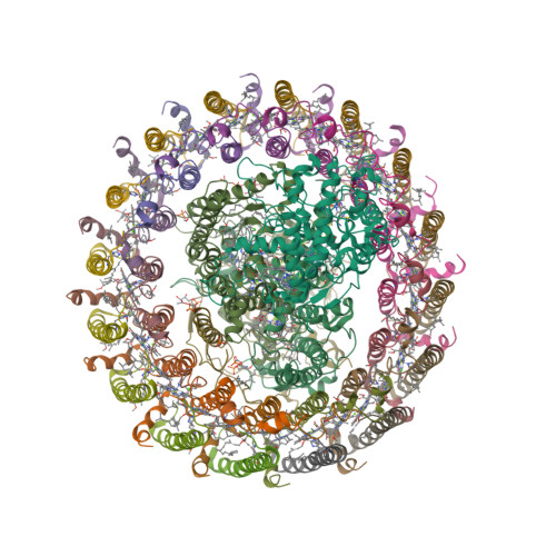
The light-harvesting core antenna (LH1) and the reaction centre (RC) of purple photosynthetic bacteria form a supramolecular complex (LH1—RC) to use sunlight energy in a highly efficient manner. Niwa et al. [1—4] report the first near-atomic structure of a LH1—RC complex, namely that of a Ca2+-bound complex from Thermochromatium tepidum. The RC is surrounded by 16 heterodimers of the LH1 αβ-subunit that form a completely closed structure. The Ca2+ ions are located at the periplasmic side of LH1. Thirty-two bacteriochlorophyll a and sixteen spirilloxanthin molecules in the LH1 ring form an elliptical assembly.

  1. Niwa, S., Yu, L.-J., Takeda, K., Hirano, Y., Kawakami, T., Wang-Otomo, Z.-Y. and Miki, K. (2014) Structure of the LH1—RC complex from Thermochromatium tepidum at 3.0 Å. Nature 508, 228—232.
  2. PDB:3WMM
  3. PDB:4V8K

Tuesday, November 26, 2013

Magnetochrome-containing iron oxidase MamP

Magnetotactic bacteria (MTB) are a diverse group of prokaryotes that have a singular ability to align with geomagnetic field lines. This ability is due to special organelles called magnetosomes. Magnetosomes are composed of single-magnetic-domain nanocrystals of magnetite [Fe(II)Fe(III)2O4] or greigite [Fe(II)Fe(III)2S4] embedded in biological membrane.

“Magnetochrome” is a name proposed in 2012 by Marina Siponen and co-authors for a cytochrome domain conserved within all known MTB and not found in any other species to date [1]. Recently, the crystal structure of the magnetosome-associated protein MamP has been solved at 1.8 Å resolution [2—4]. The minimal unit of MamP is a dimer. Each monomer consists of a PDZ domain fused to two magnetochrome domains. It was also shown in an in vitro mineralisation experiment that MamP functions as an iron oxidase mediating the iron(III) ferrihydrite production from iron(II) [2]:

4Fe2+ + 7H2O → 2Fe2O3·H2O + 12H+ + 4e
  1. Siponen, M.I., Adryanczyk, G., Ginet, N., Arnoux, P. and Pignol, D. (2012) Magnetochrome: a c-type cytochrome domain specific to magnetotatic bacteria. Biochemical Society Transactions 40, 1319—1323.
  2. Siponen, M.I., Legrand, P., Widdrat, M., Jones, S.R., Zhang, W.-J., Chang, M.C.Y., Faivre, D., Arnoux, P. and Pignol, D. (2013) Structural insight into magnetochrome-mediated magnetite biomineralization. Nature 502, 681—684.
  3. PDB:4JJ0
  4. PDB:4JJ3

Saturday, October 26, 2013

The first viral cytochrome b5

A unicellular green alga Ostreococcus tauri is the smallest (less than 1 μm in diameter) free-living eukaryote yet described. Viruses that can infect high-light and low-light adapted strains of O. tauri have been isolated and their genomes sequenced. Interestingly, low-light-strain infecting virus (OtV-2) differ from the high-light-strain infecting viruses by encoding a potential cytochrome b5 [1]. This protein was cloned, biochemically characterised and its three-dimensional structure resolved [2, 3].

The absorption spectra of oxidised and reduced recombinant OtV-2 haemoprotein are almost identical to those of purified human cytochrome b5.

Absorbance spectra of purified recombinant human cytochrome b5 and OtV-2_201.

It was also shown that the protein can substitute for yeast cytochrome b5 in the CYP51-mediated sterol 14α-demethylation. Structurally, the viral protein is similar to other known cytochromes b5 but lacks a hydrophobic C-terminal anchor. Thus, the first virally encoded cytochrome b5 is also the first cytosolic cytochrome b5 characterised. However, the physiological role of viral cytochrome b5 remains unknown.

  1. UniProt:E4WM77
  2. Reid, E.L., Weynberg, K.D., Love, J., Isupov, M.N., Littlechild, J.A., Wilson, W.H., Kelly, S.L., Lamb, D.C. and Allen, M.J. (2013) Functional and structural characterisation of a viral cytochrome b5. FEBS Letters, 587, 3633—3639.
  3. PDB:4B8N

A structural representation of the OtV-2 cytochrome b5 (OtV-2_201) protein shown as a ribbon diagram.

Saturday, August 31, 2013

Metallomics and the Cell

This volume, edited by Lucia Banci, is probably the first real book on metallomics. The table of contents looks very promising, and judging from those bits that I am able to access, I’d love to say that is is a great book... But honestly I can’t. The days when I could persuade the library to purchase (for me) a book, however expensive, are long gone. At the Springer website, eBook is priced at €142.79 and hardcover costs €181.85. You can buy them slightly cheaper from Amazon ($175.82 and $227.05, respectively.)

The book is dedicated to Ivano Bertini, who sadly passed away last year. I was lucky enough to meet the man himself on a few occasions. Ivano was a formidable scientist and one of the most colourful figures of bioinorganic chemistry and structural biology.

Metallomics and the Cell
Metal Ions in Life Sciences, vol. 12
Lucia Banci, Editor

  1. Banci, L. and Bertini, I. Metallomics and the cell: some definitions and general comments, pp. 1—13.
  2. Penner-Hahn, J.E. Technologies for detecting metals in single cells, pp. 15—40.
  3. Clausen, M.J.V. and Poulsen, H. Sodium/potassium homeostasis in the cell, pp. 41—67.
  4. Romani, A.M.P. Magnesium homeostasis in mammalian cells, pp. 69—118.
  5. Brini, M., Calì, T., Ottolini, D. and Carafoli, E. Intracellular calcium homeostasis and signaling, pp. 119—168.
  6. Roth, J., Ponzoni, S. and Aschner, M. Manganese homeostasis and transport, pp. 169—201.
  7. Andrews, S., Norton, I., Salunkhe, A.S., Goodluck, H., Aly, W.S.M., Mourad-Agha, H. and Cornelis, P. Control of iron metabolism in bacteria, pp. 203—239.
  8. Dlouhy, A.C. and Outten, C.E. The iron metallome in eukaryotic organisms, pp. 241—278.
  9. Benson, D.R. and Rivera, M. Heme uptake and metabolism in bacteria, pp. 279—332.
  10. Cracan, V. and Banerjee, R. Cobalt and corrinoid transport and biochemistry, pp. 333—374.
  11. Sydor, A.M. and Zamble, D.B. Nickel metallomics: general themes guiding nickel homeostasis, pp. 375—416.
  12. Rensing, C. and McDevitt, S.F. The copper metallome in prokaryotic cells, 417—450.
  13. Vest, K.E., Hashemi, H.F. and Cobine, P.A. The copper metallome in eukaryotic cells, pp. 451—478.
  14. Maret, W. Zinc and the zinc proteome, pp. 479—501.
  15. Mendel, R.R. Metabolism of molybdenum, pp. 503—528.
  16. Gladyshev, V.N. and Zhang, Y. Comparative genomics analysis of the metallomes, pp. 529—580.

Wednesday, July 24, 2013

Crystal structure of latex oxygenase RoxA

To date, two types of enzymes that are responsible for primary attack of polyisoprene in rubber-degrading microorganisms have been identified [1]. One is the latex clearing protein (Lcp), first isolated from Streptomyces sp., which does not have any metal ions or cofactors [2]. The other is the rubber oxygenase RoxA of Xanthomonas sp., a dihaem c-type cytochrome that cleaves cis-1,4-polyisoprene, the main constituent of natural rubber, to 12-oxo-4,8-dimethyltrideca-4,8-diene-1-al [3, 4]. The crystal structure of RoxA, solved at 1.8 Å resolution, was released today [5].

Latex Oxygenase RoxA @ PDB
  1. Birke, J., Hambsch, N., Schmitt, G., Altenbuchner, J. and Jendrossek, D. (2012) Phe317 is essential for rubber oxygenase RoxA activity. Applied and Environmental Microbiology 78, 7876—7883.
  2. Rose, K., Tenberge, K.B. and Steinbüchel, A. (2005) Identification and characterization of genes from Streptomyces sp. strain K30 responsible for clear zone formation on natural rubber latex and poly(cis-1,4-isoprene) rubber degradation. Biomacromolecules 6, 180—188.
  3. Braaz, R., Fischer, P. and Jendrossek, D. (2004) Novel type of heme-dependent oxygenase catalyzes oxidative cleavage of rubber (poly-cis-1,4-isoprene). Applied and Environmental Microbiology 70, 7388—7395.
  4. Schmitt, G., Seiffert, G., Kroneck, P.M.H., Braaz, R. and Jendrossek, D. (2010) Spectroscopic properties of rubber oxygenase RoxA from Xanthomonas sp., a new type of dihaem dioxygenase. Microbiology 156, 2537—2548.
  5. PDB:4B2N

Wednesday, May 15, 2013

Aldosterone synthase structures

Earlier this year [1], the crystal structures of human aldosterone synthase (CYP11B2) were solved in complex with a substrate 11-deoxycorticosterone [2] and an inhibitor fadrozole [3].

  1. Strushkevich, N., Gilep, A.A., Shen, L., Arrowsmith, C.H., Edwards, A.M., Usanov, S.A. and Park, H.-W. (2013) Structural insights into aldosterone synthase substrate specificity and targeted inhibition. Molecular Endocrinology 27, 315—324.
  2. PDB:4DVQ
  3. PDB:4FDH

Friday, March 22, 2013

You’re in CD spectroscopy

One day, idly browsing the web (as usual), I came across this:

I disagree with an unknown (to me) co-author of Antoine de Saint-Exupéry. For one thing, you don’t have to be “in organic chemistry” to recognise a reaction coordinate diagram. For another, 25 or so years ago my first reaction (that is, if I never read Le Petit Prince) would be: “Hey dude, your CD spectrum is upside down”. The fact is, I am still alive, so my life then was far from being over.

Isn’t the Web great? Nowadays I don’t have to venture to the library and sift through the J. Biol. Chem.’s and J. Mol. Biol.’s. (Even if I wanted, there is no library like that in Fuerteventura.) I can get the CD spectra online and for free in the Protein Circular Dichroism Data Bank [1]. Better still, using DichroMatch I can find spectra that are similar to my query [2]. (I just checked: it works!) Here’s how the CD spectrum of a typical α-helical protein (such as haemoglobin) looks like (a):


(a)

So... where’s a hat? Back in 1990s, our lab had a decommissioned Jobin Yvon Mark IV dichrograph, which, as I understand now, was an excellent machine. The haemoglobin spectrum would look more or less like this (b):


(b)

Neither equipment nor our samples allowed us to collect spectra below 200 nm, therefore most of the spectrum was in the negative ellipticity region. We did not really need to go below 200 nm: we were mostly monitoring ellipticity at 222 nm as a function of temperature or concentration of guanidinium chloride or other denaturing agents.

Mind you, not all proteins have this inverted hat region in their CD spectra. For example, ferredoxin (c), rubredoxin (d) or immunoglobulin G (e):


(c)


(d)


(e)

In the 21st century, protein X-ray crystallography became very much a routine technique. Once you solve the structure, there’s no mystery left. On the contrary, the CD spectra are as beautiful and enigmatic as star spectra. They still need an intelligent interpreter. They tell the story and in the same time keep the secret. I think the little prince would appreciate them.

  1. Whitmore, L., Woollett, B., Miles, A.J., Klose, D.P., Janes, R.W. and Wallace, B.A. (2011) PCDDB: the protein circular dichroism data bank, a repository for circular dichroism spectral and metadata. Nucleic Acids Research 39, D480—D486.
  2. Klose, D.P., Wallace, B.A. and Janes, R.W. (2012) DichroMatch: a website for similarity searching of circular dichroism spectra. Nucleic Acids Research 40, W547—W552.
Thursday, February 28, 2013

The first CYP1A1 structure

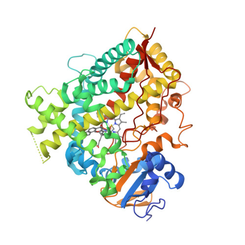
CYP1A1 was one of the first P450 enzymes to be characterised and, as its name indicates, holds the first place in the systematic nomenclature of P450s [1]. However, it was not until last year that the first crystal structure of human CYP1A1 in complex with α-naphthoflavone has been determined at 2.6 Å resolution [2]. The structure [3] is released this week.

  1. Nebert, D.W., Nelson, D.R., Coon, M.J., Estabrook, R.W., Feyereisen, R., Fujii-Kuriyama, Y., Gonzalez, F.J., Guengerich, F.P., Gunsalus, I.C., Johnson, E.F., Loper, J.C., Sato, R., Waterman, M.R. and Waxman, D.J. (1991) The P450 superfamily: update on new sequences, gene mapping, and recommended nomenclature. DNA Cell Biol. 10, 1—14.
  2. Walsh, A.A., Szklarz, G.D. and Scott, E.E. (2012) Cytochrome P450 1A1 structure and utility in predicting drug and xenobiotic metabolism. Proceedings of the 19th International Symposium on Microsomes and Drug Oxidations and 12th European ISSX Meeting, Noordwijk, The Netherlands, 17—21 June 2012.
  3. PDB:4I8V

Monday, December 24, 2012

Macromolecular Xmas decorations

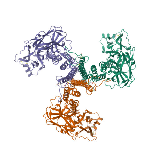
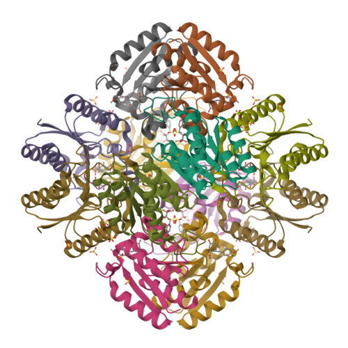
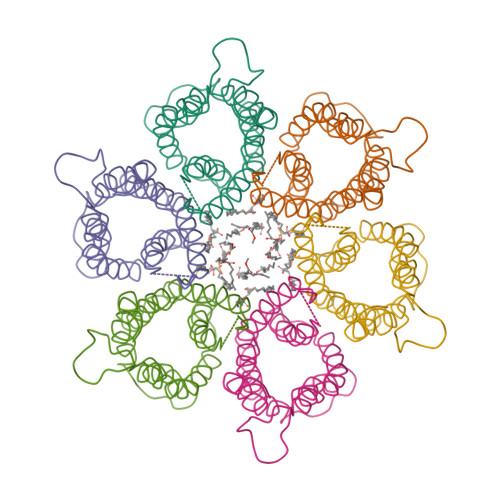
This year I don’t have a Christmas tree. (There aren’t many in Fuerteventura.) But I’ve got some models from this week’s new PDB structures [1—5] which look Christmassy enough to decorate my blog. Merry Christmas and a happy New Year everybody!


  1. Hui, F., Scheib, U., Hu, Y., Sommer, R.J., Aroian, R.V. and Ghosh, P. (2012) Structure and glycolipid binding properties of the nematicidal protein Cry5B. Biochemistry 51, 9911—9921.
  2. Lence, E., Tizón, L., Otero, J.M., Peón, A., Prazeres, V.F.V., Llamas-Saiz, A.L., Fox, G.C., van Raaij, M.J., Lamb, H., Hawkins, A.R. and González-Bello, C. (2013) Mechanistic basis of the inhibition of type II dehydroquinase by (2S)- and (2R)-2-benzyl-3-dehydroquinic acids. ACS Chem. Biol. 8, 568—577.
  3. Strugatsky, D., McNulty, R., Munson, K., Chen, C.-K., Soltis, S.M., Sachs, G. and Luecke, H. (2013) Structure of the proton-gated urea channel from the gastric pathogen Helicobacter pylori. Nature 493, 255—258.
  4. Tang, Q., Gao, P., Liu, Y.-P., Gao, A., An, X.-M., Liu, S., Yan, X.-X. and Liang, D.-C. (2012) RecOR complex including RecR N-N dimer and RecO monomer displays a high affinity for ssDNA. Nucleic Acids Res. 40, 11115—11125.
  5. Ziervogel, B.K. and Roux, B. (2013) The binding of antibiotics in OmpF porin. Structure 21, 76—87.