Showing posts with label cryo-electron microscopy. Show all posts

Friday, December 25, 2015

Human Apaf-1 apoptosome at 3.8 Å

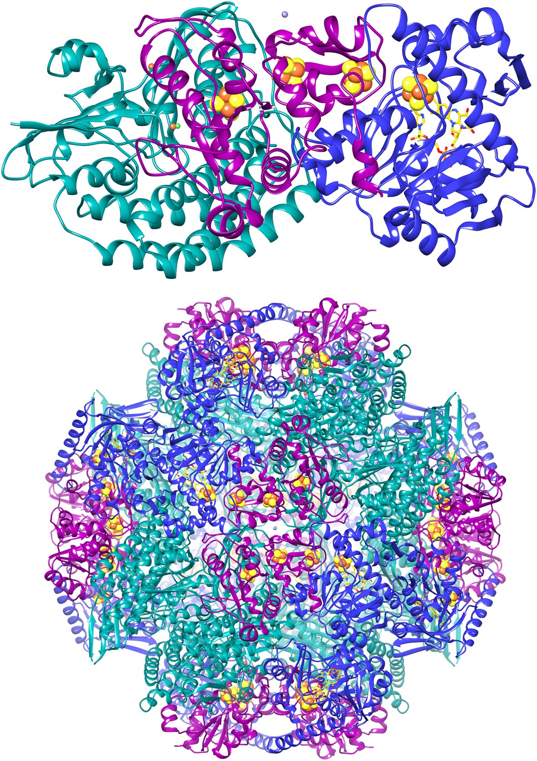
The apoptotic protease-activating factor 1 (Apaf-1) exists in normal cells as an autoinhibited monomer. Upon binding to cytochrome c and dATP, Apaf-1 forms a heptameric complex known as the apoptosome. Zhou et al. report an atomic structure of an intact human Apaf-1 apoptosome at 3.8 Å resolution determined by single-particle, cryo-electron microscopy [1, 2].

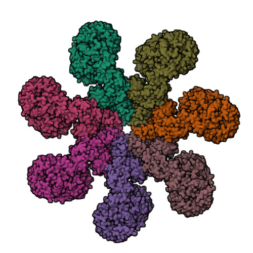
  1. Zhou, M., Li, Y., Hu, Q., Bai, X.-c., Huang, W., Yan, C., Scheres, S.H.W. and Shi, Y. (2015) Atomic structure of the apoptosome: mechanism of cytochrome c- and dATP-mediated activation of Apaf-1. Genes & Development 29, 2349—2361.
  2. PDB:3JBT

Thursday, July 31, 2014

F420-reducing [NiFe]-hydrogenase at 1.7 Å

The F420-reducing [NiFe]-hydrogenase (FrhABG; EC 1.12.98.1) catalyses the reversible redox reaction between coenzyme F420 and H2. FrhABG is a group 3 [NiFe]-hydrogenase with a dodecameric quaternary structure recently revealed by high-resolution cryo-electron microscopy [1]. Vitt et al. report the crystal structure of FrhABG from Methanothermobacter marburgensis at 1.7 Å resolution [2, 3] and compare it with the structures of group 1 [NiFe]-hydrogenases, the only previously structurally characterised group.

  1. Allegretti, M., Mills, D.J., McMullan, G., Kühlbrandt, W. and Vonck, J. (2014) Atomic model of the F420-reducing [NiFe] hydrogenase by electron cryo-microscopy using a direct electron detector. eLife 3, e01963.
  2. Vitt, S., Ma, K., Warkentin, E., Moll, J., Pierik, A.J., Shima, S. and Ermler, U. (2014) The F420-reducing [NiFe]-hydrogenase complex from Methanothermobacter marburgensis, the first X-ray structure of a group 3 family member. J. Mol. Biol. 426, 2813—2826.
  3. PDB:4OMF