Showing posts with label photosynthesis. Show all posts

Saturday, February 27, 2016

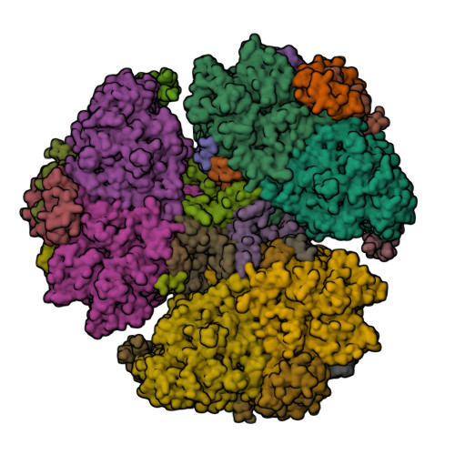
First eukaryotic photosystem II solved at 2.76 Å

The water-splitting reaction of photosynthesis is catalysed by photosystem II (PSII), a large protein complex located in thylakoid membranes of organisms ranging from cyanobacteria to higher plants [1]. During the last 15 years, a number of crystal structures of PSII from cyanobacteria have been solved. However, no structures of PSII from eukaryots have been reported until now, partly due to the instability of eukaryotic PSII upon isolation. Ago et al. [2] solved the structure of PSII from a red alga Cyanidium caldarium at 2.76 Å resolution [3]. This PSII contains four extrinsic proteins, including the three subunits found in cyanobacterial PSII and the fourth subunit PsbQ' homologous to the PsbQ protein of green algae and higher plants. Furthermore, two novel trans-membrane helices were found in the algal PSII which are not present in cyanobacterial PSII.

  1. Shen, J.-R. (2015) The structure of photosystem II and the mechanism of water oxidation in photosynthesis. Annual Review of Plant Biology 66, 23—48.
  2. Ago, H., Adachi, H., Umena, Y., Tashiro, T., Kawakami, K., Kamiya, N., Tian, L., Han, G., Kuang, T., Liu, Z., Wang, F., Zou, H., Enami, I., Miyano, M. and Shen, J.-R. (2016) Novel features of eukaryotic photosystem II revealed by its crystal structure analysis from a red alga. J. Biol. Chem. 291, 5676—5687.
  3. PDB:4YUU

Monday, June 30, 2014

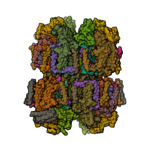
Phycocyanin against Alzheimer’s?

Could a light-harvesting protein phycocyanin be used as a novel drug against Alzheimer’s disease (AD) [1, 2]?

In the present study, intact hexameric phycocyanin was isolated and crystallized from the cyanobacterium Leptolyngbya sp. N62DM, and the structure was solved to a resolution of 2.6 Å. Molecular docking studies show that the phycocyanin αβ-dimer interacts with the enzyme β-secretase, which catalyzes the proteolysis of the amyloid precursor protein to form plaques. The molecular docking studies suggest that the interaction between phycocyanin and β-secretase is energetically more favorable than previously reported inhibitor-β-secretase interactions. Transgenic Caenorhabditis elegans worms, with a genotype to serve as an AD-model, were significantly protected by phycocyanin. Therefore, the present study provides a novel structure-based molecular mechanism of phycocyanin-mediated therapy against AD.
  1. Singh, N.K., Hasan, S.S., Kumar, J., Raj, I., Pathan, A.A., Parmar, A., Shakil, S., Gourinath, S. and Madamwar D. (2014) Crystal structure and interaction of phycocyanin with β-secretase: A putative therapy for Alzheimer's disease. CNS Neurol. Disord. Drug Targets 13, 691—698.
  2. PDB:4L1E

Tuesday, April 22, 2014

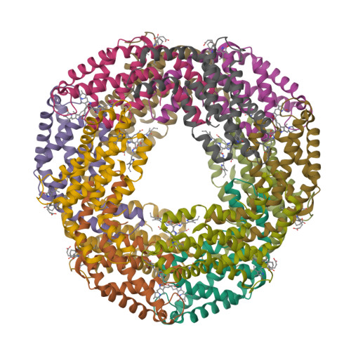
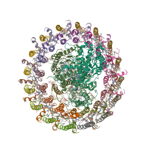
Thermochromatium tepidum LH1—RC complex at 3.0 Å

The light-harvesting core antenna (LH1) and the reaction centre (RC) of purple photosynthetic bacteria form a supramolecular complex (LH1—RC) to use sunlight energy in a highly efficient manner. Niwa et al. [1—4] report the first near-atomic structure of a LH1—RC complex, namely that of a Ca2+-bound complex from Thermochromatium tepidum. The RC is surrounded by 16 heterodimers of the LH1 αβ-subunit that form a completely closed structure. The Ca2+ ions are located at the periplasmic side of LH1. Thirty-two bacteriochlorophyll a and sixteen spirilloxanthin molecules in the LH1 ring form an elliptical assembly.

  1. Niwa, S., Yu, L.-J., Takeda, K., Hirano, Y., Kawakami, T., Wang-Otomo, Z.-Y. and Miki, K. (2014) Structure of the LH1—RC complex from Thermochromatium tepidum at 3.0 Å. Nature 508, 228—232.
  2. PDB:3WMM
  3. PDB:4V8K

Monday, March 21, 2011

Femtosecond X-ray nanocrystallography of PSI

It is a well known fact (to those who know it well) that in order to obtain a decent diffraction pattern one has to grow a decent-size crystal first. Well, that is about to change. The PDB entry enigmatically named “femtosecond X-ray protein nanocrystallography” [1] in fact contains the structure of the photosystem I (PSI) from Thermosynechococcus elongatus solved by this new method [2]. In this work, more that 3 million diffraction patterns were collected from really small PSI crystals (from ~200  nm to 2  μm in size) illuminated by the new femtosecond X-ray laser, the Linac Coherent Light Source in Stanford. According to the authors,

We mitigate the problem of radiation damage in crystallography by using pulses briefer than the timescale of most damage processes.
  1. PDB:3PCQ
  2. Chapman, H.N., Fromme, P., Barty, A. et al. (2011) Femtosecond X-ray protein nanocrystallography. Nature 470, 73—77.

Thursday, February 04, 2010

The rise and fall of the Zinc World

I like the way the journals such as Biology Direct now publish reviewers’ comments and authors’ responses together with a final version of the paper. The fact that reviewers’ names are made public ensure that reviewers are properly acknowledged as well as share some responsibility for releasing another pointless paper into the wild. The discussion is often more interesting than a paper. Take the couple of recent highly speculative articles on Zinc world and origin of life [1, 2]. Even though I myself would not recommend any of these manuscripts for publication (luckily nobody asked me), I am glad that these were eventually published, because I really enjoyed reading the reviews — and, occasionally, the authors’ responses.

Reviewer 4: Last but not least I find the last sentence of the paper rather revealing: what could the aesthetics of minerals to do with a scientific argument on the origin of life?

Author’s response: Aesthetic criteria are of great importance in scientific research <...> For example, my initial opposition to the idea of abiogenesis at the floor of the Hadean ocean, when I first heard about it, was purely aesthetic. I simply did not like the idea of the origin of life being in complete darkness.
Similarly, it could have been stated “I simply liked the idea of using a single type of metal cation to fulfill all life’s metal needs”. The authors acknowledge the need for transition metal ions for origin of life but argue that the zinc is preferred to other transition metals because it is not redox-active. (According to modern view, zinc is not a transition element at all — why to bring transition metals in the first place?) For example, both papers contain the statement that “iron, unlike zinc, is redox-active”. Wait a minute. Is it bad? Since zinc is not redox-active, it cannot be used as a redox cofactor, therefore there won’t be any oxidoreductases utilising zinc. But fear not; apparently, the authors think that NAD(P)H, FAD and FMN evolved before primitive life forms learned how to use Fe2+ ions safely.

Incidentally, the only danger of redox-active iron mentioned in these papers seems to be the “harmful hydroxyl radicals”. I would not worry much about them though because I don’t think they were the main hazard in largely anoxygenic environment. In general, conditions on Earth at the time were rather harsh. You would’t go outside without an oxygen mask and a very thick (a few inches?) layer of sunscreen. Now add, on top of that, ZnS-catalysed photosynthetic production of formaldehyde [2, equation 1]... yep, sounds a plausible enough way to kick off that life thing.

  1. Mulkidjanian, A.Y. (2009) On the origin of life in the Zinc world: 1. Photosynthesizing, porous edifices built of hydrothermally precipitated zinc sulfide as cradles of life on Earth. Biology Direct 4, 26.
  2. Mulkidjanian, A.Y. and Galperin, M.Y. (2009) On the origin of life in the Zinc world. 2. Validation of the hypothesis on the photosynthesizing zinc sulfide edifices as cradles of life on Earth. Biology Direct 4, 27.