Showing posts with label biomacromolecules. Show all posts

Saturday, January 23, 2021

Chains and rings

After hours spent looking in my books and searching the internet, I came to the conclusion that chemists talk about chains and rings without explaining what they mean. The only definition I found so far, viz. that of Gold Book, is specific for polymers and seems to be too complex to be used in general chemical nomenclature:

The whole or part of a macromolecule, an oligomer molecule or a block, comprising a linear or branched sequence of constitutional units between two boundary constitutional units, each of which may be either an end-group, a branch point or an otherwise-designated characteristic feature of the macromolecule.
(1)

On the other hand, general dictionary definitions of (chemical) chains are not precise enough. For example, Collins English Dictionary defines chain (chemistry) as

two or more atoms or groups bonded together so that the configuration of the resulting molecule, ion, or radical resembles a chain.
(2)

whereas Merriam-Webster says that it is

a number of atoms or chemical groups united like links in a chain.
(3)

So chain (chemistry) is like a chain. Is it?

Saturday, February 27, 2016

First eukaryotic photosystem II solved at 2.76 Å

The water-splitting reaction of photosynthesis is catalysed by photosystem II (PSII), a large protein complex located in thylakoid membranes of organisms ranging from cyanobacteria to higher plants [1]. During the last 15 years, a number of crystal structures of PSII from cyanobacteria have been solved. However, no structures of PSII from eukaryots have been reported until now, partly due to the instability of eukaryotic PSII upon isolation. Ago et al. [2] solved the structure of PSII from a red alga Cyanidium caldarium at 2.76 Å resolution [3]. This PSII contains four extrinsic proteins, including the three subunits found in cyanobacterial PSII and the fourth subunit PsbQ' homologous to the PsbQ protein of green algae and higher plants. Furthermore, two novel trans-membrane helices were found in the algal PSII which are not present in cyanobacterial PSII.

  1. Shen, J.-R. (2015) The structure of photosystem II and the mechanism of water oxidation in photosynthesis. Annual Review of Plant Biology 66, 23—48.
  2. Ago, H., Adachi, H., Umena, Y., Tashiro, T., Kawakami, K., Kamiya, N., Tian, L., Han, G., Kuang, T., Liu, Z., Wang, F., Zou, H., Enami, I., Miyano, M. and Shen, J.-R. (2016) Novel features of eukaryotic photosystem II revealed by its crystal structure analysis from a red alga. J. Biol. Chem. 291, 5676—5687.
  3. PDB:4YUU

Saturday, January 30, 2016

Crystal structure of the DNAzyme 9DB1

The first ever crystal structure of a deoxyribozyme has been solved at 2.8 Å resolution [1—3]. The work by researchers from Max Planck Institute for Biophysical Chemistry (Göttingen, Germany) also sheds light on a difference in catalytic mechanism of ribozymes and deoxyribozymes [4]:

Ribozymes use RNA’s 2´-hydroxyl groups, which are absent in DNA, for structural interactions or directly for catalysis. The new structure shows why the lack of these groups doesn’t diminish the catalytic activity of DNAzymes. The missing hydroxyls make DNA’s sugar-phosphate backbone more flexible, allowing acrobatic conformations that compensate for the absent hydroxyls in DNAzymes.

  1. Ponce-Salvatierra, A., Wawrzyniak-Turek, K., Steuerwald, U., Höbartner, C. and Pena, V. (2016) Crystal structure of a DNA catalyst. Nature 529, 231—234.
  2. PDB:5CKK
  3. PDB:5CKI
  4. Borman, S. (2016) After two decades of trying, scientists report first crystal structure of a DNAzyme. Chemical & Engineering News 94, issue 2, p. 3.

Friday, December 25, 2015

Human Apaf-1 apoptosome at 3.8 Å

The apoptotic protease-activating factor 1 (Apaf-1) exists in normal cells as an autoinhibited monomer. Upon binding to cytochrome c and dATP, Apaf-1 forms a heptameric complex known as the apoptosome. Zhou et al. report an atomic structure of an intact human Apaf-1 apoptosome at 3.8 Å resolution determined by single-particle, cryo-electron microscopy [1, 2].

  1. Zhou, M., Li, Y., Hu, Q., Bai, X.-c., Huang, W., Yan, C., Scheres, S.H.W. and Shi, Y. (2015) Atomic structure of the apoptosome: mechanism of cytochrome c- and dATP-mediated activation of Apaf-1. Genes & Development 29, 2349—2361.
  2. PDB:3JBT

Thursday, November 19, 2015

A magnetic protein biocompass?

A team of scientists from Peking University report a putative magnetic receptor (MagR) protein in Drosophila, CG8198 [1]. MagR binds an iron—sulphur cluster and interacts with photoreceptor cryptochrome (Cry) proteins to form a multimeric magnetosensing rod-like complex. Assemblies of these rods were observed orienting themselves in a weak magnetic field. Qin et al. speculate that these structures may function like compasses in living organisms, although the mechanism of magnetoreception in vivo remains a mystery [2].

A complete Cry/MagR magnetosensor protein complex structure model with 10 Crys helically binding to the rod-like MagR polymer consisting of 20 MagRs.
  1. Qin, S., Yin, H., Yang, C., Dou, Y., Liu, Z., Zhang, P., Yu, H., Huang, Y., Feng, J., Hao, J., Hao, J., Deng, L., Yan, X., Dong, X., Zhao, Z., Jiang, T., Wang, H.-W., Luo, S.-J. and Xie, C. (2016) A magnetic protein biocompass. Nature Materials 15, 217–226.
  2. Cyranoski, D. (2015) Long-sought biological compass discovered: Protein complex offers explanation for how animals sense Earth’s magnetic pull. Nature 527, 283–284.

Sunday, September 27, 2015

Nitrite binding modes in CuNIR

Fukuda and Inoue [1] have determined the crystal structure of the C135A mutant of thermostable copper nitrite reductase (CuNIR) from Geobacillus thermodenitrificans in complex with nitrite to 1.55 Å resolution. Interestingly, this high-temperature (320 K) structure [2] displays a near-bidentate binding mode of nitrite distinct from a monodentate mode in a cryogenic structure [3]:

To our knowledge, this is the first case in which the difference in substrate binding modes between cryogenic and high-temperature structures has been visualized by crystallography.

The copper site geometries are given in Table 1.

Table 1 (adapted from [4])

Cu—Ligand Distances (Å) 3X1N (320 K) 3WKP (100 K)
I. Type 1 Cu—residue distances
T1Cu—H95Nδ1 2.08 2.14
T1Cu—H143Nδ1 2.01 1.96
T1Cu—M148Sδ 2.13 2.07
II. Type 2 Cu—residue distances
T2Cu—H100Nε2 2.07 1.96
T2Cu—H134Nε2 2.00 1.95
T2Cu—H294Nε2 1.98 2.01
T2Cu—water 2.02 n/a
III. Type 2 Cu—nitrite distances
T2Cu—Oproximal 2.13 1.97
T2Cu—N 2.21 2.85
T2Cu—Odistal 2.52 3.41
  1. Fukuda, Y. and Inoue, T. (2015) High-temperature and high-resolution crystallography of thermostable copper nitrite reductase. Chemical Communications 51, 6532—6535.
  2. PDB:3X1N
  3. PDB:3WKP
  4. Fukuda, Y. and Inoue, T. (2015) High-temperature and high-resolution crystallography of thermostable copper nitrite reductase. Electronic Supplementary Material.

Wednesday, July 24, 2013

Crystal structure of latex oxygenase RoxA

To date, two types of enzymes that are responsible for primary attack of polyisoprene in rubber-degrading microorganisms have been identified [1]. One is the latex clearing protein (Lcp), first isolated from Streptomyces sp., which does not have any metal ions or cofactors [2]. The other is the rubber oxygenase RoxA of Xanthomonas sp., a dihaem c-type cytochrome that cleaves cis-1,4-polyisoprene, the main constituent of natural rubber, to 12-oxo-4,8-dimethyltrideca-4,8-diene-1-al [3, 4]. The crystal structure of RoxA, solved at 1.8 Å resolution, was released today [5].

Latex Oxygenase RoxA @ PDB
  1. Birke, J., Hambsch, N., Schmitt, G., Altenbuchner, J. and Jendrossek, D. (2012) Phe317 is essential for rubber oxygenase RoxA activity. Applied and Environmental Microbiology 78, 7876—7883.
  2. Rose, K., Tenberge, K.B. and Steinbüchel, A. (2005) Identification and characterization of genes from Streptomyces sp. strain K30 responsible for clear zone formation on natural rubber latex and poly(cis-1,4-isoprene) rubber degradation. Biomacromolecules 6, 180—188.
  3. Braaz, R., Fischer, P. and Jendrossek, D. (2004) Novel type of heme-dependent oxygenase catalyzes oxidative cleavage of rubber (poly-cis-1,4-isoprene). Applied and Environmental Microbiology 70, 7388—7395.
  4. Schmitt, G., Seiffert, G., Kroneck, P.M.H., Braaz, R. and Jendrossek, D. (2010) Spectroscopic properties of rubber oxygenase RoxA from Xanthomonas sp., a new type of dihaem dioxygenase. Microbiology 156, 2537—2548.
  5. PDB:4B2N

Friday, March 22, 2013

You’re in CD spectroscopy

One day, idly browsing the web (as usual), I came across this:

I disagree with an unknown (to me) co-author of Antoine de Saint-Exupéry. For one thing, you don’t have to be “in organic chemistry” to recognise a reaction coordinate diagram. For another, 25 or so years ago my first reaction (that is, if I never read Le Petit Prince) would be: “Hey dude, your CD spectrum is upside down”. The fact is, I am still alive, so my life then was far from being over.

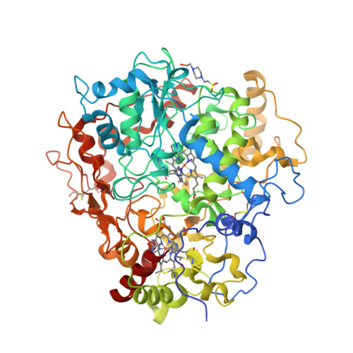
Isn’t the Web great? Nowadays I don’t have to venture to the library and sift through the J. Biol. Chem.’s and J. Mol. Biol.’s. (Even if I wanted, there is no library like that in Fuerteventura.) I can get the CD spectra online and for free in the Protein Circular Dichroism Data Bank [1]. Better still, using DichroMatch I can find spectra that are similar to my query [2]. (I just checked: it works!) Here’s how the CD spectrum of a typical α-helical protein (such as haemoglobin) looks like (a):


(a)

So... where’s a hat? Back in 1990s, our lab had a decommissioned Jobin Yvon Mark IV dichrograph, which, as I understand now, was an excellent machine. The haemoglobin spectrum would look more or less like this (b):


(b)

Neither equipment nor our samples allowed us to collect spectra below 200 nm, therefore most of the spectrum was in the negative ellipticity region. We did not really need to go below 200 nm: we were mostly monitoring ellipticity at 222 nm as a function of temperature or concentration of guanidinium chloride or other denaturing agents.

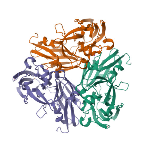
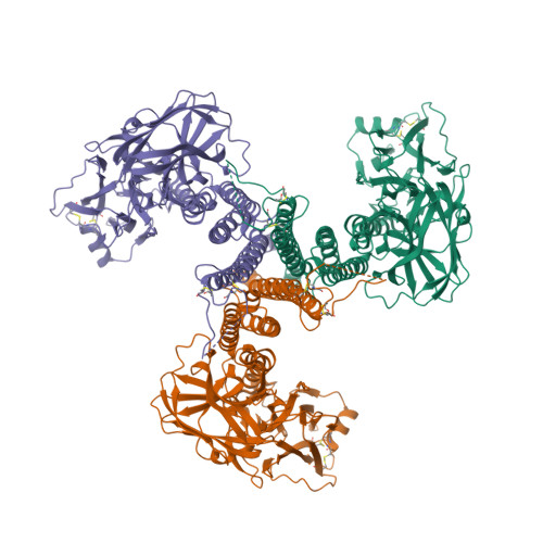
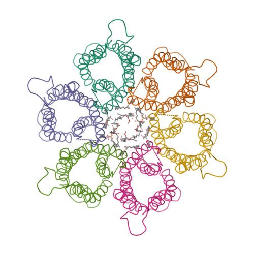
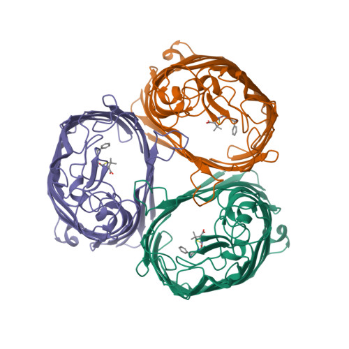
Mind you, not all proteins have this inverted hat region in their CD spectra. For example, ferredoxin (c), rubredoxin (d) or immunoglobulin G (e):


(c)


(d)


(e)

In the 21st century, protein X-ray crystallography became very much a routine technique. Once you solve the structure, there’s no mystery left. On the contrary, the CD spectra are as beautiful and enigmatic as star spectra. They still need an intelligent interpreter. They tell the story and in the same time keep the secret. I think the little prince would appreciate them.

  1. Whitmore, L., Woollett, B., Miles, A.J., Klose, D.P., Janes, R.W. and Wallace, B.A. (2011) PCDDB: the protein circular dichroism data bank, a repository for circular dichroism spectral and metadata. Nucleic Acids Research 39, D480—D486.
  2. Klose, D.P., Wallace, B.A. and Janes, R.W. (2012) DichroMatch: a website for similarity searching of circular dichroism spectra. Nucleic Acids Research 40, W547—W552.

Monday, December 24, 2012

Macromolecular Xmas decorations

This year I don’t have a Christmas tree. (There aren’t many in Fuerteventura.) But I’ve got some models from this week’s new PDB structures [1—5] which look Christmassy enough to decorate my blog. Merry Christmas and a happy New Year everybody!


  1. Hui, F., Scheib, U., Hu, Y., Sommer, R.J., Aroian, R.V. and Ghosh, P. (2012) Structure and glycolipid binding properties of the nematicidal protein Cry5B. Biochemistry 51, 9911—9921.
  2. Lence, E., Tizón, L., Otero, J.M., Peón, A., Prazeres, V.F.V., Llamas-Saiz, A.L., Fox, G.C., van Raaij, M.J., Lamb, H., Hawkins, A.R. and González-Bello, C. (2013) Mechanistic basis of the inhibition of type II dehydroquinase by (2S)- and (2R)-2-benzyl-3-dehydroquinic acids. ACS Chem. Biol. 8, 568—577.
  3. Strugatsky, D., McNulty, R., Munson, K., Chen, C.-K., Soltis, S.M., Sachs, G. and Luecke, H. (2013) Structure of the proton-gated urea channel from the gastric pathogen Helicobacter pylori. Nature 493, 255—258.
  4. Tang, Q., Gao, P., Liu, Y.-P., Gao, A., An, X.-M., Liu, S., Yan, X.-X. and Liang, D.-C. (2012) RecOR complex including RecR N-N dimer and RecO monomer displays a high affinity for ssDNA. Nucleic Acids Res. 40, 11115—11125.
  5. Ziervogel, B.K. and Roux, B. (2013) The binding of antibiotics in OmpF porin. Structure 21, 76—87.

Monday, September 21, 2009

Sulfimide bond in collagen IV

The recent paper in Science describes the sulfilimine (sulfimide, in IUPACese) bond, “not previously found in biomolecules”, identified in collagen. The bond (>S=N–) cross-links methionine and hydroxylysine residues of adjoining protomers.