Wednesday, September 30, 2009

Uranyl-binding protein

“Uranyl ion” is the traditional name of dioxidouranium(2+), [UO2]2+. According to Wikipedia, [UO2]2+ is “the most common species encountered in the aqueous chemistry of uranium”. Wegner et al. designed a uranyl-selective DNA-binding protein using the template of NikR, a nickel-dependent transcriptional repressor from E. coli. The binding site of wild-type NikR was modified by a series of mutations (Val72Ser, His76Asp and Cys95Asp) to introduce extra hard equatorial ligands to favour binding of [UO2]2+.

Wild-type NikR binds to its promoter DNA in the presence of Ni2+ ions, and a number of other divalent metal ions such as Cu2+, Zn2+, Co2+, Mn2+, and Cd2+ can also induce protein-DNA complex formation. However, NikR does not bind to DNA in the presence of 50 μM UO22+. The <triple> mutant NikR′ binds to DNA neither in the absence of metal ions nor in the presence of Ni2+ ions, but it forms a protein-DNA complex in the presence of UO22+. In comparison to NikR, the metal selectivity of NikR′ has been altered. Experiments with other metal ions show that the mutant protein only forms the protein-DNA complex in the presence of the uranyl cation while Ni2+, Zn2+, Co2+, Cu2+, Cd2+, Mn2+, and Fe2+ ions do not result in any observable complex formation. Attempts to load NikR with uranyl or NikR′ with Ni2+ did not yield any observable metal binding. Thus, this mutant NikR′ shows a uranyl-specific DNA-binding ability.

Sunday, September 27, 2009

Coloured coins

In contrast to gold-, silver- and copper-coloured coins (whatever metal they are made of), simply “coloured coins” sport the colours not usually associated with coinage metals. Or so I think, because so far I could not find any definitive guide to coloured coins. Wikipedia mentions them in an article on 
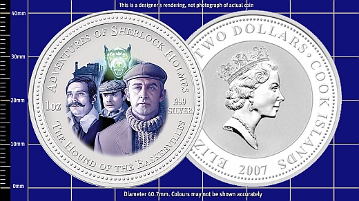
commemorative coins. I got interested in the subject since I discovered that the Sherlock Holmes Silver Coins produced by the New Zealand Mint (apparently, the legal tender of the Cook Islands) are graced by the images of Sherlock Holmes and Doctor Watson from Soviet-era films, such as The Hound of the Baskervilles.


The Tony Clayton’s website contains a very useful list of metals used in coins and medals. In particular, I have learned that niobium is used as coinage metal. In 2003, Münze Österreich pioneered the use of niobium for coin manufacturing, issuing a bimetallic €25 coin. According to this review,
The colouring <of the niobium insert> is made by a so called anodic oxidation of the material. With this treatment, by electrochemical processing a very thin niobium oxide layer is formed under controlled conditions. By refraction of light in the oxide layer so called interference colours are created which gives the colouring of the niobium. Depending on the processing parameters, the thickness of the oxide layer can be very well controlled, and gives the niobium its noble appearance. Depending on the thickness of the layer different colours are producible.
For instance, Latvian bimetallic Coin of Time (struck by Münze Österreich) consists of beautiful blue niobium centre enclosed in an outer silver ring. The obverse of the coin features the heraldic rose and the tiny Gothic script letters and standing for Heinrich Rose (1795—1864), discoverer of niobium.

Latvia-Coin of Time (obverse).gifLatvia-Coin of Time (reverse).gif

Wednesday, September 23, 2009

Magnetic monopoles in spin ice

Although the magnetic monopoles were postulated by Paul Dirac in 1931, the existence of these particles remains an open problem. This article surveys the recent breakthrough discoveries concerning magnetic monopoles within spin ice materials, dysprosium titanate (Dy2Ti2O7) and holmium titanate (Ho2Ti2O7).

Monday, September 21, 2009

Sulfimide bond in collagen IV

The recent paper in Science describes the sulfilimine (sulfimide, in IUPACese) bond, “not previously found in biomolecules”, identified in collagen. The bond (>S=N–) cross-links methionine and hydroxylysine residues of adjoining protomers.

Wednesday, September 16, 2009

Stereochemistry of digitonin

Following the call to the community from Antony Williams, I indulged in some chemical drawing. I did redraw structure 1 from this paper from scratch to get (a). This is very much like structure of digitonin in ChEBI (b), except for methyl group at C-20 which goes up in (a).

20beta-digitonin
(a)
20alpha-digitonin
(b)

Muhr et al. wrote:

With our investigations, it was possible for the first time to confirm beyond all doubt the structure suggested by Tschesche and Wulff for digitonin by means of modern NMR techniques, and to assign all proton and carbon resonances.

Now I was not able to get to the full text of Tschesche and Wulff, but at least their abstract contains the German name “3[β-D-Glucopyranosyl(I)(1→3Galakt.II)-β-D-galaktopyranosyl(II)(1→2Gluc.III)-β-D-xylopyranosyl(1→3Gluc.III)-β-D-glucopyranosyl(III) (1→4Galakt.IV)-β-D-galaktopyranosyl(IV)(1→3-Digitog.)]5α,20βF,25α Spirostantriol(2α,3β,15β)”, which kind of confirms 20β configuration. (The default configuration of spirostan is 20α.)

I guess this still does not answer what the “correct” structure of digitonin is. All we can say that Muhr et al. reported the structure (a).

Friday, September 11, 2009

Charge-shift bonding

Here’s something one doesn’t see in a chemistry textbook. The recent perspective paper in Nature Chemistry deals with a distinct class of electron-pair bonding called “charge-shift” (CS) bonding, which exists alongside classical covalent and ionic bonding. And in not-so exotic molecules.

<A> striking example is the difference between H2 and F2; two homonuclear bonds that by all criteria should be classified as covalent bonds, but exhibit fundamental differences. Consider the energy curves (Fig. 1) of the two bonds calculated recently. Figure 1a shows that the H—H bond is indeed covalent; its covalent structure accounts for most of the bonding energy (relative to the ‘exact’ curve). By contrast, for the F—F bond in Fig. 1b, the covalent structure is entirely repulsive, and what determines the bonding energy and the equilibrium distance is the covalent–ionic mixing. This mixing leads to a resonance energy stabilization, which we have termed the ‘charge-shift resonance energy’ (RECS). Thus, despite their apparent similarity, the two bonds are very different; whereas the H—H bond is a true covalent bond, the F—F bond is a CS bond that is completely determined by the RECS quantity.

No less striking example is so-called inverted C—C bond in [1.1.1]propellane (described in a paper from the same group of authors), which “closely resembles the single bond of difluorine”.

Tuesday, September 01, 2009

Antarcticite

Antarcticite is a mineral form of calcium dichloride hexahydrate. It was first discovered in Don Juan Pond in Antarctica, which is probably the saltiest (47% w/v) body of water on earth. Looking at crystal structure of antarcticite (below), one can see that both name “calcium dichloride hexahydrate” and formula CaCl2·6H2O are misleading, for there are two kinds of water in it. The structure comprises the alternating layers of (i) trigonal planar triaquacalcium(2+) ions and (ii) water and chloride ions. I suppose it should be named “triaquacalcium dichloride—water (1/3)” or “triaquacalcium dichloride trihydrate”, with formula [Ca(OH2)3]Cl2·3H2O.

anarcticite