Tuesday, September 23, 2014

Pseudomonas fluorescens PhoX

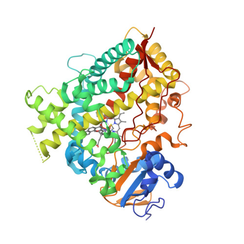
Alkaline phosphatases (EC 3.1.3.1) occur widely in nature and are found in all three domains of life [1]. The Escherichia coli PhoA enzyme has been extensively studied whereas PhoX family of alkaline phosphatases are only minimally characterised and show no sequence similarity to other phosphotransfer enzymes. Yong et al. [2] determined high-resolution crystal structures for native PhoX from Pseudomonas fluorescens [3] and for its complexes with phosphate [4], a nonhydrolysable ATP analogue adenosine-5′-[β,γ-methylene]triphosphate (AMP-PCP) [5], and the putative transition-state mimic vanadate [6]. The active site contains two antiferromagnetically coupled ferric ions (Fe3+), three calcium ions (Ca2+), and an oxo group bridging one Ca2+ and two Fe3+ ions.

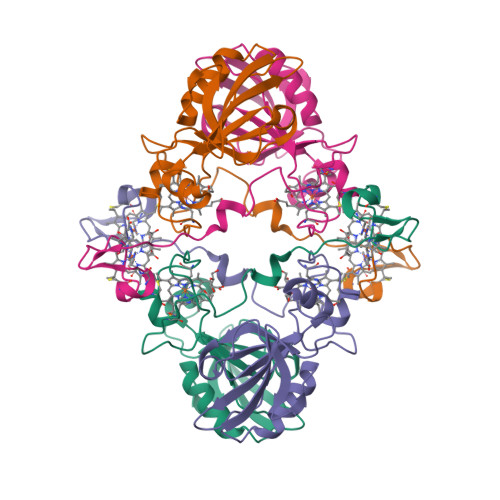
Cartoon representation of P. fluorescens PhoX crystal structure.
The PhoX active site containing bound phosphate [1, Fig. 2c].
A model for the catalytic mechanism of PhoX [1, Fig. 3d].
The transition state is indicated with the double dagger (‡) symbol.
  1. Millán, J.L. (2006) Alkaline Phosphatases: Structure, substrate specificity and functional relatedness to other members of a large superfamily of enzymes. Purinergic Signalling 2, 335–341.
  2. Yong, S.C., Roversi, P., Lillington, J., Rodriguez, F., Krehenbrink, M., Zeldin, O.B., Garman, E.F., Lea, S.M. and Berks, B.C. (2014) A complex iron-calcium cofactor catalyzing phosphotransfer chemistry. Science 345, 1170—1173.
  3. PDB:4A9V
  4. PDB:4ALF
  5. PDB:4AMF
  6. PDB:3ZWU

Tuesday, August 26, 2014

Ogres are not like cakes

I was intrigued by the article in New Scientist which starts with the question, “Do you speak chemistry?” [1]. So much that I asked my friend to send me the original paper [2] authored by the Bartosz Grzybowski group of Northwestern University in Evanston, Illinois. It is a curious reading.

Don’t get me wrong. I have nothing against the analogies. I love the analogies. If the linguistic analogy works for chemistry, it’s fine by me. As long as everybody understands that it is just an analogy.

The authors try to “demonstrate that a natural language such as English and organic chemistry have the same structure in terms of the frequency of, respectively, text fragments and molecular fragments”. How do they do that? They start by looking at the maximum common substrings (MCS) found in 100 sentences randomly chosen from English Wikipedia.

Perhaps not surprisingly, the most common fragment of the sentences is “e”, followed by “a” and “o”.
That is surprising to me though, considering that only “a” is a word in English. I wouldn’t be surprised if it happened to be Spanish Wikipedia. Are the authors talking about letter frequency per chance? But the “top three” letters in English (from most to least common) are known to be E, T, A while in Spanish they are E, A, O. Anyway, they show that the distribution of the fragments, whatever they are, follows the power law. Then they show that the distribution of the common molecular fragments, derived from the corpus of organic molecules, also follows the power law. Big deal: so do the earthquake magnitudes, populations of cities and stock market crashes [3]. Cadeddu et al. do not seem to be bothered with that at all:
We have just shown that there exists a set of molecular fragments with which organic molecules can be described akin to a language.
So far so bad; whether you are a linguist, a computational chemist or an organic chemist, both methodology and conclusions of this paper are bound to make you cringe. So, my immediate reaction was to dismiss it altogether. Ogres are not like cakes. Organic molecules are not like a language. End of story.

But could it be that I am missing something? On the one hand, the language of chemistry — whether we are talking trivial names, systematic names, or graphical diagrams — is very much like any other language: a system of communication. On the other hand, the molecules themselves are not. Unless they are the information macromolecules. The message encoded in a single DNA molecule can be very much abstracted from its chemical structure. Without any doubt, genetic code is a communication system, therefore it is a language, although not man-made.

It’s interesting that the authors view organic molecules as “sentences” rather than “words”; the latter would be the nomenclaturist’s approach. I guess it depends on your taste, or language preferences. Most systematic chemical names look alien in English but would fit rather nicely in German or Finnish. I personally view any chemical name as a noun phrase describing a corresponding molecular entity; a molecular entity itself is not a noun phrase. However, in natural languages, there rarely is a confusion regarding the boundaries of a word:

a word is the smallest element that may be uttered in isolation with semantic or pragmatic content (with literal or practical meaning).
On the contrary, Grzybowski’s “words” are the molecular fragments which do not exist in isolation. It is also worth noting that in the world of biopolymers, say nucleic acids, each monomer (as complex as any of Grzybowski’s “sentences”), is often represented as a letter, while an entire bacterial genome (still a single DNA molecule) could be considered a War and Peace (or Crime and Punishment).

Cadeddu et al. further claim that linguistic approach identifies the symmetry/repeat units in molecules such as α-cyclodextrin and porphyrin:

We emphasize that this is not a small feat given we have not even considered any (x, y, z) coordinates of the atoms making up these molecules and performed no linear-algebra analyses to find symmetries which, incidentally, can be a computationally intensive procedure involving manipulation of matrices.
I find this modest remark regarding the size of the “feat” within the body of a scientific article in a respectable journal really cute. Are the authors even aware that there are chemical similarity/substructure search engines? You don’t need atomic coordinates to identify the fragments with the same connectivity.

Which brings me to the final point. What is the “chemical linguistics” anyway? If the “words” of chemistry, as postulated in [2], are nothing else but molecular fragments, or substructures, then the chemoinformaticians were doing the substructure search of chemical databases for donkey’s years without knowing that it is called chemical linguistics. I am aware of completely different use of this term in a sense “mining of natural language texts for chemical information” [4, 5]. This latter use is well-established and I think applying the name “chemical linguistics” to unrelated area will only confuse everybody.

  1. Aron, J. (2014) Language of chemistry is unveiled by molecular make-up. New Scientist no. 2981, p. 8.
  2. Cadeddu, A., Wylie, E.K., Jurczak, J., Wampler-Doty, M. and Grzybowski, B.A. (2014) Organic chemistry as a language and the implications of chemical linguistics for structural and retrosynthetic analyses. Angewandte Chemie 126, 8246—8250.
  3. Buchanan, M. (2000) Ubiquity, Weidenfeld & Nicolson, London.
  4. Goebels, L., Grotz, H., Lawson, A.L., Roller, S. and Wisniewski, J. (2005) Method and software for extracting chemical data. Patent DE 102005020083 A1.
  5. Day, N.E., Corbett, P.T. and Murray-Rust, P. (2007) Semantic chemical publishing. ACS National Meeting #233, Chicago.

Thursday, July 31, 2014

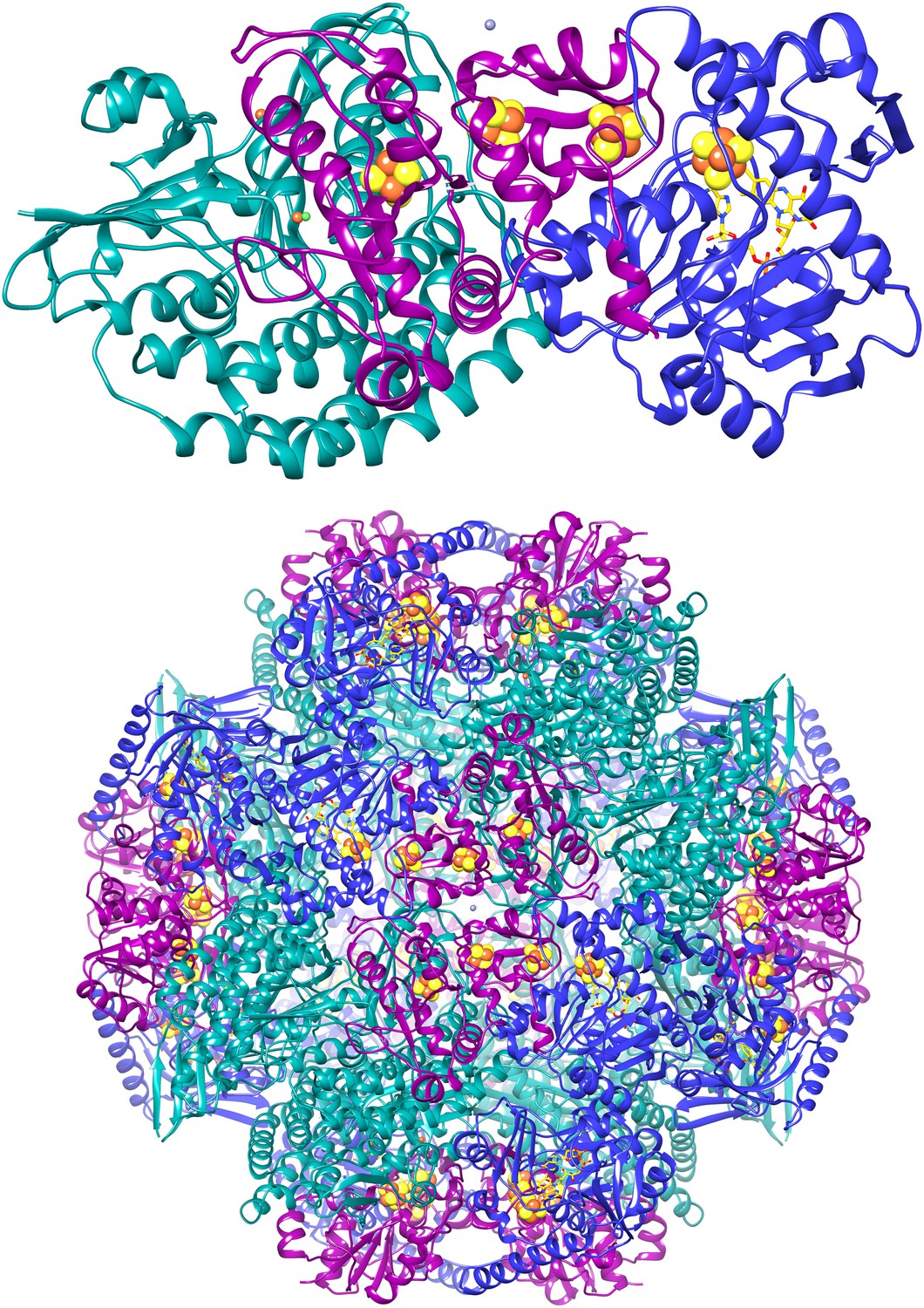
F420-reducing [NiFe]-hydrogenase at 1.7 Å

The F420-reducing [NiFe]-hydrogenase (FrhABG; EC 1.12.98.1) catalyses the reversible redox reaction between coenzyme F420 and H2. FrhABG is a group 3 [NiFe]-hydrogenase with a dodecameric quaternary structure recently revealed by high-resolution cryo-electron microscopy [1]. Vitt et al. report the crystal structure of FrhABG from Methanothermobacter marburgensis at 1.7 Å resolution [2, 3] and compare it with the structures of group 1 [NiFe]-hydrogenases, the only previously structurally characterised group.

  1. Allegretti, M., Mills, D.J., McMullan, G., Kühlbrandt, W. and Vonck, J. (2014) Atomic model of the F420-reducing [NiFe] hydrogenase by electron cryo-microscopy using a direct electron detector. eLife 3, e01963.
  2. Vitt, S., Ma, K., Warkentin, E., Moll, J., Pierik, A.J., Shima, S. and Ermler, U. (2014) The F420-reducing [NiFe]-hydrogenase complex from Methanothermobacter marburgensis, the first X-ray structure of a group 3 family member. J. Mol. Biol. 426, 2813—2826.
  3. PDB:4OMF

Monday, June 30, 2014

Phycocyanin against Alzheimer’s?

Could a light-harvesting protein phycocyanin be used as a novel drug against Alzheimer’s disease (AD) [1, 2]?

In the present study, intact hexameric phycocyanin was isolated and crystallized from the cyanobacterium Leptolyngbya sp. N62DM, and the structure was solved to a resolution of 2.6 Å. Molecular docking studies show that the phycocyanin αβ-dimer interacts with the enzyme β-secretase, which catalyzes the proteolysis of the amyloid precursor protein to form plaques. The molecular docking studies suggest that the interaction between phycocyanin and β-secretase is energetically more favorable than previously reported inhibitor-β-secretase interactions. Transgenic Caenorhabditis elegans worms, with a genotype to serve as an AD-model, were significantly protected by phycocyanin. Therefore, the present study provides a novel structure-based molecular mechanism of phycocyanin-mediated therapy against AD.
  1. Singh, N.K., Hasan, S.S., Kumar, J., Raj, I., Pathan, A.A., Parmar, A., Shakil, S., Gourinath, S. and Madamwar D. (2014) Crystal structure and interaction of phycocyanin with β-secretase: A putative therapy for Alzheimer's disease. CNS Neurol. Disord. Drug Targets 13, 691—698.
  2. PDB:4L1E

Saturday, May 24, 2014

[Fe3S4] ferredoxin from Rhodopseudomonas palustris

The crystal structure of a novel [Fe3S4] ferredoxin associated with CYP194A4 from Rhodopseudomonas palustris has been solved at 2.15 Å resolution [1—3]. The ferredoxin, HaPuxC, contains an atypical CXXHXXC(X)nCP iron-sulphur cluster-binding motif. HaPuxC is the first P450 electron-transfer partner of this type to be structurally characterised.

  1. Zhang, T., Zhang, A., Bell, S.G., Wong, L.-L. and Zhou, W. (2014) The structure of a novel electron-transfer ferredoxin from Rhodopseudomonas palustris HaA2 which contains a histidine residue in its iron-sulfur cluster-binding motif. Acta Crystallographica D70, 1453—1464.
  2. PDB:4ID8
  3. PDB:4OV1

Thursday, May 01, 2014

Tetracalcium octachromium(3+) strontium octacarbonate hexadecahydroxide sulfate pentaicosahydrate

The Polar Bear peninsula in Western Australia is one of the many places on this planet I never heard before. The reason I mention it now is that a new mineral named putnisite was discovered there, and this mineral caused a bit of a stir recently, for being “completely unique and unrelated to anything”. In fact, if you Google “Polar Bear peninsula”, all you find is putnisite.

In 2007, specimens of an unknown mineral forming purple crystals (a) were collected at the Polar Bear peninsula while prospecting for nickel and gold. The specimens were eventually forwarded to Peter Elliott, a research associate with the South Australian Museum, for examination.

(a)

Elliott et al. [2] report the composition and crystal structure of this unique mineral, named in honour of mineralogists Christine and Andrew Putnis of the Institut für Mineralogie, Universtität Münster, Germany. The compositional name for putnisite I come up with is “tetracalcium octachromium(3+) strontium octacarbonate hexadecahydroxide sulfate pentaicosahydrate”. Curiously, Mindat and Mineralienatlas give the molecular formula containing only 23 molecules of water.

(b)

The crystal structure (b) was determined from single-crystal X-ray diffraction data. Cr(OH)4O2 octahedra (red) link by edge-sharing to form an eight-membered ring. At the centre of each ring lies a decacoordinated Sr2+ cation (purple). The rings are decorated by carbonate triangles (green), each of which links by corner-sharing to two Cr(OH)4O2 octahedra. Rings are linked by Ca(H2O)4O4 polyhedra (blue) to form a sheet parallel to the (100) plane. Adjacent sheets are joined along the [100] direction by corner-sharing sulfate tetrahedra (yellow).

  1. Mills, R. (2014) New mineral shows nature’s infinite variability. Phys.org.
  2. Elliott, P., Giester, G., Rowe, R. and Pring, A. (2014) Putnisite, SrCa4Cr3+8(CO3)8SO4(OH)16·25H2O, a new mineral from Western Australia: description and crystal structure. Mineralogical Magazine 78, 131—144.

Tuesday, April 22, 2014

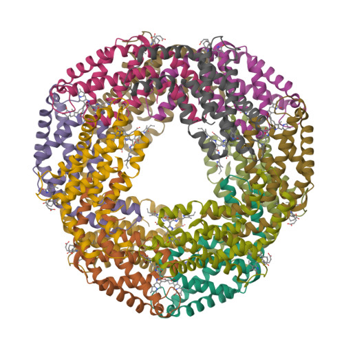
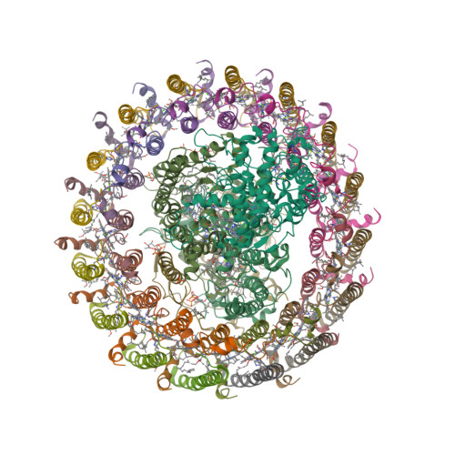
Thermochromatium tepidum LH1—RC complex at 3.0 Å

The light-harvesting core antenna (LH1) and the reaction centre (RC) of purple photosynthetic bacteria form a supramolecular complex (LH1—RC) to use sunlight energy in a highly efficient manner. Niwa et al. [1—4] report the first near-atomic structure of a LH1—RC complex, namely that of a Ca2+-bound complex from Thermochromatium tepidum. The RC is surrounded by 16 heterodimers of the LH1 αβ-subunit that form a completely closed structure. The Ca2+ ions are located at the periplasmic side of LH1. Thirty-two bacteriochlorophyll a and sixteen spirilloxanthin molecules in the LH1 ring form an elliptical assembly.

  1. Niwa, S., Yu, L.-J., Takeda, K., Hirano, Y., Kawakami, T., Wang-Otomo, Z.-Y. and Miki, K. (2014) Structure of the LH1—RC complex from Thermochromatium tepidum at 3.0 Å. Nature 508, 228—232.
  2. PDB:3WMM
  3. PDB:4V8K

Thursday, February 27, 2014

Smell of pine vs climate change

That’s right: the smell of pine trees from boreal forests could mitigate the global warming — provided that the global warming doesn’t kill the forests first [1]. The volatile organic compounds (VOCs), responsible for the smell of pine, react with atmospheric oxygen to form aerosols. These aerosols scatter solar radiation and also act as cloud condensation nuclei, thereby affecting the Earth’s radiation balance. An international group including researchers from Finland, Germany, Denmark and USA has discovered a direct pathway leading from several biogenic VOCs, such as monoterpenes, to the formation of extremely low-volatility vapours (ELVOCs) [2].

These vapours form at significant mass yield in the gas phase and condense irreversibly onto aerosol surfaces to produce secondary organic aerosol, helping to explain the discrepancy between the observed atmospheric burden of secondary organic aerosol and that reported by many model studies. We further demonstrate how these low-volatility vapours can enhance, or even dominate, the formation and growth of aerosol particles over forested regions, providing a missing link between biogenic VOCs and their conversion to aerosol particles.

Structures of the main VOCs studied by Ehn et al. [2]

The air was sampled in Hyytiälä, Finland and the chamber experiments were conducted at the Jülich Research Centre in Germany.

  1. McGrath, M. Smell of forest pine can limit climate change. BBC News, 26 February 2014.
  2. Ehn, M., Thornton, J.A., Kleist, E., Sipilä, M., Junninen, J., Pullinen, I., Springer, M., Rubach, F., Tillmann, R., Lee, B., Lopez-Hilfiker, F., Andres, S., Acir, I.-H., Rissanen, M., Jokinen, T., Schobesberger, S., Kangasluoma, J., Kontkanen, J., Nieminen, T., Kurtén, T., Nielsen, L.B., Jørgensen, S., Kjaergaard, H.G., Canagaratna, M., Dal Maso, M.D., Berndt, T., Petäjä, T., Wahner, A., Veli-Matti Kerminen, V.-M., Kulmala, M., Worsnop, D.R., Wildt, J. and Mentel, T.F. (2014) A large source of low-volatility secondary organic aerosol. Nature 506, 476—479.

Saturday, January 18, 2014

Sodium chloride revisited

Everybody knows that the formula of sodium chloride is NaCl. Right? Right. But recently, the team of Artem Oganov at Stony Brook University have shown that there are other stable types of crystalline sodium chloride. They have predicted several thermodynamically stable compounds: Na3Cl, Na2Cl, Na3Cl2, NaCl3, and NaCl7. Moreover, by utilising high-pressure techniques, they synthesised cubic and orthorhombic NaCl3 and two-dimensional tetragonal Na3Cl [1, 2].

NaCl3 (space group Pm3n)
Na3Cl (space group P4/mmm)

“One of these materials — Na3Cl — has a fascinating structure”, says Oganov. “It is comprised of layers of NaCl and layers of pure sodium. The NaCl layers act as insulators; the pure sodium layers conduct electricity” [3].

  1. Zhang, W., Oganov, A.R., Goncharov, A.F., Zhu, Q., Boulfelfel, S.E., Lyakhov, A.O., Stavrou, E., Somayazulu, M., Prakapenka, V.B. and Konôpková, Z. (2013) Unexpected stable stoichiometries of sodium chlorides. Science 342, 1502—1505; arXiv:1310.7674v1
  2. Ibáñez Insa, J. (2013) Reformulating table salt under pressure. Science 342, 1459—1460.
  3. SBU team discovers new compounds that challenge the foundation of chemistry. Stony Brook University Newsroom, December 19, 2013.

Tuesday, November 26, 2013

Magnetochrome-containing iron oxidase MamP

Magnetotactic bacteria (MTB) are a diverse group of prokaryotes that have a singular ability to align with geomagnetic field lines. This ability is due to special organelles called magnetosomes. Magnetosomes are composed of single-magnetic-domain nanocrystals of magnetite [Fe(II)Fe(III)2O4] or greigite [Fe(II)Fe(III)2S4] embedded in biological membrane.

“Magnetochrome” is a name proposed in 2012 by Marina Siponen and co-authors for a cytochrome domain conserved within all known MTB and not found in any other species to date [1]. Recently, the crystal structure of the magnetosome-associated protein MamP has been solved at 1.8 Å resolution [2—4]. The minimal unit of MamP is a dimer. Each monomer consists of a PDZ domain fused to two magnetochrome domains. It was also shown in an in vitro mineralisation experiment that MamP functions as an iron oxidase mediating the iron(III) ferrihydrite production from iron(II) [2]:

4Fe2+ + 7H2O → 2Fe2O3·H2O + 12H+ + 4e–
  1. Siponen, M.I., Adryanczyk, G., Ginet, N., Arnoux, P. and Pignol, D. (2012) Magnetochrome: a c-type cytochrome domain specific to magnetotatic bacteria. Biochemical Society Transactions 40, 1319—1323.
  2. Siponen, M.I., Legrand, P., Widdrat, M., Jones, S.R., Zhang, W.-J., Chang, M.C.Y., Faivre, D., Arnoux, P. and Pignol, D. (2013) Structural insight into magnetochrome-mediated magnetite biomineralization. Nature 502, 681—684.
  3. PDB:4JJ0
  4. PDB:4JJ3

Saturday, October 26, 2013

The first viral cytochrome b5

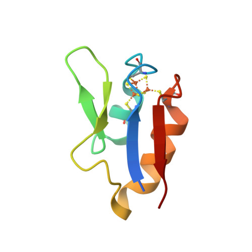
A unicellular green alga Ostreococcus tauri is the smallest (less than 1 μm in diameter) free-living eukaryote yet described. Viruses that can infect high-light and low-light adapted strains of O. tauri have been isolated and their genomes sequenced. Interestingly, low-light-strain infecting virus (OtV-2) differ from the high-light-strain infecting viruses by encoding a potential cytochrome b5 [1]. This protein was cloned, biochemically characterised and its three-dimensional structure resolved [2, 3].

The absorption spectra of oxidised and reduced recombinant OtV-2 haemoprotein are almost identical to those of purified human cytochrome b5.

Absorbance spectra of purified recombinant human cytochrome b5 and OtV-2_201.

It was also shown that the protein can substitute for yeast cytochrome b5 in the CYP51-mediated sterol 14α-demethylation. Structurally, the viral protein is similar to other known cytochromes b5 but lacks a hydrophobic C-terminal anchor. Thus, the first virally encoded cytochrome b5 is also the first cytosolic cytochrome b5 characterised. However, the physiological role of viral cytochrome b5 remains unknown.

  1. UniProt:E4WM77
  2. Reid, E.L., Weynberg, K.D., Love, J., Isupov, M.N., Littlechild, J.A., Wilson, W.H., Kelly, S.L., Lamb, D.C. and Allen, M.J. (2013) Functional and structural characterisation of a viral cytochrome b5. FEBS Letters, 587, 3633—3639.
  3. PDB:4B8N

A structural representation of the OtV-2 cytochrome b5 (OtV-2_201) protein shown as a ribbon diagram.

Thursday, September 26, 2013

Ochre, a great universal

From Architecture of First Societies: A Global Perspective by Mark Jarzombek:

Known scientifically as hematite, ochre is a reddish iron-containing rock that was used as a coloring substance made by grinding the stone into a powder that when mixed with fat or water formed a paste. Homo erectus had already begun to work with ochre, thus securing it as a key, nonfunctional element in human life.
Ochre was a great universal of all First Societies. The Blackfeet of the American Plains referred to it as nitsisaan or “real paint” and profusely daubed it on their ceremonial garments. Its color was thought to represent the sun and the energy that permeates all things, making a person rubbed with it appear holy and powerful. Its redness and brilliance signaled supernatural potency overlapping with a range of cosmological concepts revolving around rain, fertility, hunting, and death. The nineteen-century painter George Catlin painted the Sioux worshipping at a red boulder in the open grasslands of the Great Plains.
Sioux Worshiping at the Red Boulders
George Catlin, Sioux Worshiping at the Red Boulders, 1837—1839
Ceremonial uses of ochre still exist today, such as among the Maasai in Kenya during certain initiation rituals, by Amazon tribes and by Aboriginal people in Australia.
The !Kung, who live in the Kalahari desert of Botswana and who are among the oldest of the surviving First Society people in the world, use the pigment in rituals dealing with a woman’s first menstruation. A female initiate, on emergence from seclusion, would present the women of her kin group with lumps of ochre for decorating their faces and cloaks and also for adorning the young men to protect them when out hunting.

Saturday, August 31, 2013

Metallomics and the Cell

This volume, edited by Lucia Banci, is probably the first real book on metallomics. The table of contents looks very promising, and judging from those bits that I am able to access, I’d love to say that is is a great book... But honestly I can’t. The days when I could persuade the library to purchase (for me) a book, however expensive, are long gone. At the Springer website, eBook is priced at €142.79 and hardcover costs €181.85. You can buy them slightly cheaper from Amazon ($175.82 and $227.05, respectively.)

The book is dedicated to Ivano Bertini, who sadly passed away last year. I was lucky enough to meet the man himself on a few occasions. Ivano was a formidable scientist and one of the most colourful figures of bioinorganic chemistry and structural biology.

Metallomics and the Cell
Metal Ions in Life Sciences, vol. 12
Lucia Banci, Editor

  1. Banci, L. and Bertini, I. Metallomics and the cell: some definitions and general comments, pp. 1—13.
  2. Penner-Hahn, J.E. Technologies for detecting metals in single cells, pp. 15—40.
  3. Clausen, M.J.V. and Poulsen, H. Sodium/potassium homeostasis in the cell, pp. 41—67.
  4. Romani, A.M.P. Magnesium homeostasis in mammalian cells, pp. 69—118.
  5. Brini, M., Calì, T., Ottolini, D. and Carafoli, E. Intracellular calcium homeostasis and signaling, pp. 119—168.
  6. Roth, J., Ponzoni, S. and Aschner, M. Manganese homeostasis and transport, pp. 169—201.
  7. Andrews, S., Norton, I., Salunkhe, A.S., Goodluck, H., Aly, W.S.M., Mourad-Agha, H. and Cornelis, P. Control of iron metabolism in bacteria, pp. 203—239.
  8. Dlouhy, A.C. and Outten, C.E. The iron metallome in eukaryotic organisms, pp. 241—278.
  9. Benson, D.R. and Rivera, M. Heme uptake and metabolism in bacteria, pp. 279—332.
  10. Cracan, V. and Banerjee, R. Cobalt and corrinoid transport and biochemistry, pp. 333—374.
  11. Sydor, A.M. and Zamble, D.B. Nickel metallomics: general themes guiding nickel homeostasis, pp. 375—416.
  12. Rensing, C. and McDevitt, S.F. The copper metallome in prokaryotic cells, 417—450.
  13. Vest, K.E., Hashemi, H.F. and Cobine, P.A. The copper metallome in eukaryotic cells, pp. 451—478.
  14. Maret, W. Zinc and the zinc proteome, pp. 479—501.
  15. Mendel, R.R. Metabolism of molybdenum, pp. 503—528.
  16. Gladyshev, V.N. and Zhang, Y. Comparative genomics analysis of the metallomes, pp. 529—580.

Wednesday, July 24, 2013

Crystal structure of latex oxygenase RoxA

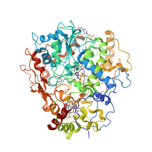
To date, two types of enzymes that are responsible for primary attack of polyisoprene in rubber-degrading microorganisms have been identified [1]. One is the latex clearing protein (Lcp), first isolated from Streptomyces sp., which does not have any metal ions or cofactors [2]. The other is the rubber oxygenase RoxA of Xanthomonas sp., a dihaem c-type cytochrome that cleaves cis-1,4-polyisoprene, the main constituent of natural rubber, to 12-oxo-4,8-dimethyltrideca-4,8-diene-1-al [3, 4]. The crystal structure of RoxA, solved at 1.8 Å resolution, was released today [5].

Latex Oxygenase RoxA @ PDB
  1. Birke, J., Hambsch, N., Schmitt, G., Altenbuchner, J. and Jendrossek, D. (2012) Phe317 is essential for rubber oxygenase RoxA activity. Applied and Environmental Microbiology 78, 7876—7883.
  2. Rose, K., Tenberge, K.B. and Steinbüchel, A. (2005) Identification and characterization of genes from Streptomyces sp. strain K30 responsible for clear zone formation on natural rubber latex and poly(cis-1,4-isoprene) rubber degradation. Biomacromolecules 6, 180—188.
  3. Braaz, R., Fischer, P. and Jendrossek, D. (2004) Novel type of heme-dependent oxygenase catalyzes oxidative cleavage of rubber (poly-cis-1,4-isoprene). Applied and Environmental Microbiology 70, 7388—7395.
  4. Schmitt, G., Seiffert, G., Kroneck, P.M.H., Braaz, R. and Jendrossek, D. (2010) Spectroscopic properties of rubber oxygenase RoxA from Xanthomonas sp., a new type of dihaem dioxygenase. Microbiology 156, 2537—2548.
  5. PDB:4B2N

Saturday, June 22, 2013

S-Adenosyl-S-carboxymethyl-L-homocysteine

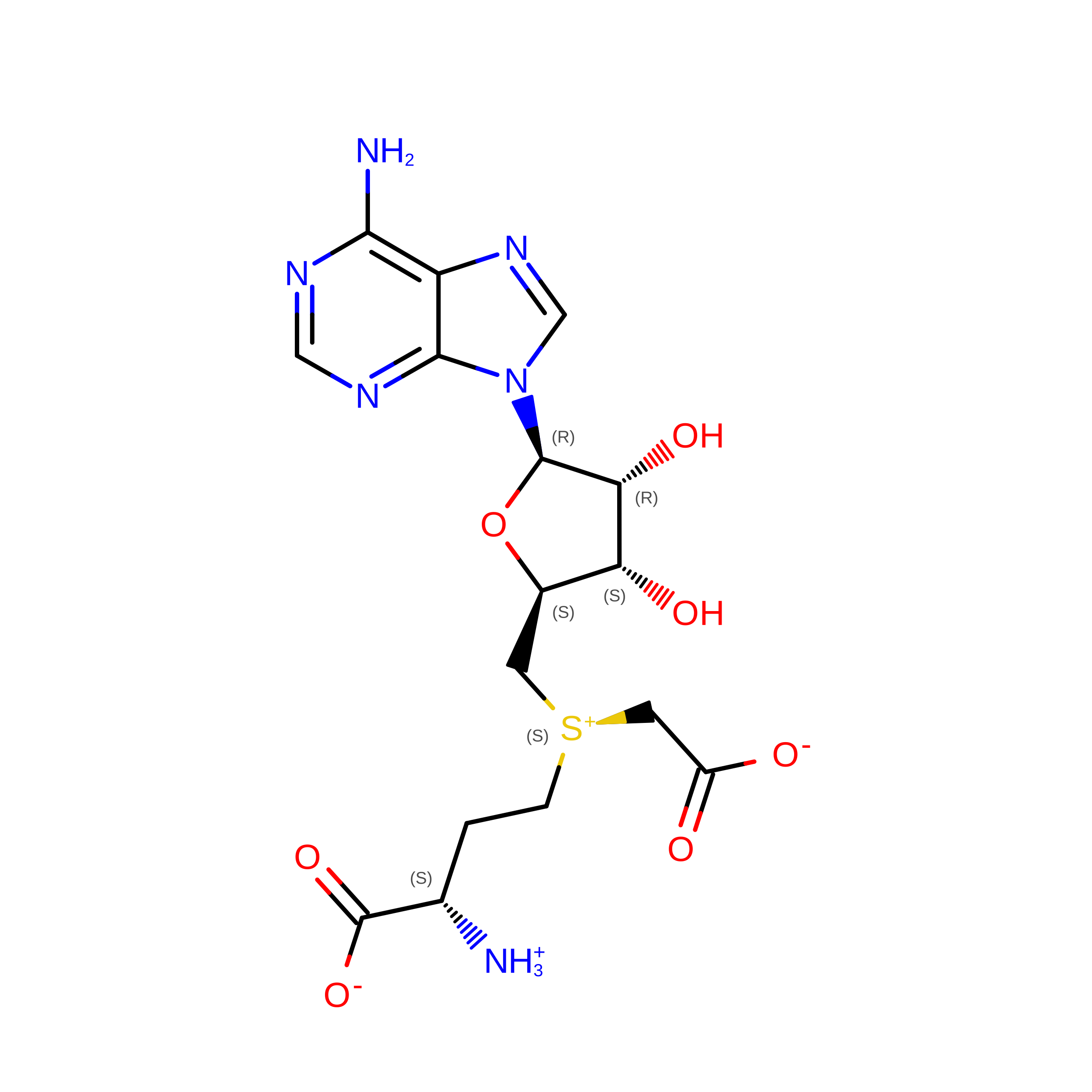
A novel cofactor is not something that is discovered every day, or even every year. So we are lucky this year. The crystal structure of a putative methyltransferase CmoA from Escherichia coli reveals the presence of [(3S)-3-amino-3-carboxypropyl]{[(2S,3S,4R,5R)-5-(6-aminopurin-9-yl)-3,4-dihydroxyoxolan-2-yl]methyl}(carboxymethyl)sulfanium, aka S-adenosyl-S-carboxymethyl-L-homocysteine, aka SCM-SAH [1—3]. Moreover, it was suggested that “a number of enzymes that have previously been annotated as SAM-dependent are in fact SCM-SAH-dependent” [1].

  1. Byrne, R.T., Whelan, F., Aller, P., Bird, L.E., Dowle, A., Lobley, C.M., Reddivari, Y., Nettleship, J.E., Owens, R.J., Antson, A.A. and Waterman, D.G. (2013) S-Adenosyl-S-carboxymethyl-L-homocysteine: a novel cofactor found in the putative tRNA-modifying enzyme CmoA. Acta Crystallographica D69, 1090—1098.
  2. PDB:4GEK
  3. PDB:4IWN

Wednesday, May 15, 2013

Aldosterone synthase structures

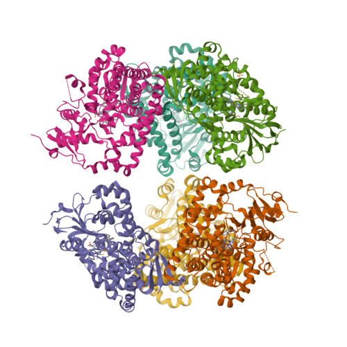
Earlier this year [1], the crystal structures of human aldosterone synthase (CYP11B2) were solved in complex with a substrate 11-deoxycorticosterone [2] and an inhibitor fadrozole [3].

  1. Strushkevich, N., Gilep, A.A., Shen, L., Arrowsmith, C.H., Edwards, A.M., Usanov, S.A. and Park, H.-W. (2013) Structural insights into aldosterone synthase substrate specificity and targeted inhibition. Molecular Endocrinology 27, 315—324.
  2. PDB:4DVQ
  3. PDB:4FDH

Sunday, April 14, 2013

Supercool

Once again, Summer is upon us. And what could be more satisfying on a hot Summer day than cold beer? I’ll tell you what: really cold beer.

In any good Spanish bar, they will serve you beer in a frozen glass, or copa fría. Here’s a pair of well-chilled beer tankards:

When you pour cold beer in copa fría, it will form ice crystals:

How much ice is formed, depends on temperature and beer strength. In general, the stronger the drink, the lower the freezing point.

On a number of occasions, I was chilling beer in the freezer. Then taking it out, opening the bottle and pouring it into the glass. There are four experimentally observed outcomes.

  1. Beer is liquid both in a bottle and in a glass
  2. Beer is liquid in a bottle but gets frozen as poured in a glass
  3. Beer gets solid in a bottle so no way to pour it in a glass
  4. Beer bottle explodes in the freezer
Scenario B is the most interesting one. (Scenario D is also interesting, but I won’t recommend it.) This is how the resulting beer slush looks like:

Now I saw a number of articles on the web where they explain this phenomenon with supercooling. I should say that I am not satisfied with this explanation. Why “supercooled” beer is not getting frozen in the bottle, even if I shake it, but forms slush once outside? When I put a bottle of (non-fizzy) rosé in a freezer, it either stays liquid (and remains liquid upon opening and pouring) or develops fine crystals of ice (which stay as they are upon opening both in a bottle and in a glass). On one occasion, a forgotten in a freezer bottle of rosé got frozen solid. (According to The Academic Wino, “the freezing point of table wine is –5 °C”, and my freezer goes down to –18 °C.)

To explain what happens, we don’t need to bring supercooling in. We just have to keep in mind that our drink is an aqueous solution. And that alcohol is only one of many solutes there. Of them, the most important are sugars and carbon monoxide. (Some beers, such as Guinness, contain dissolved dinitrogen as well as CO2.)

For dissolution to take place, the overall change of free energy should be negative, but the heat may be either absorbed or released. The dissolving of sugar in water is an endothermic process. The increase in temperature results in an increase in solubility. The reverse process, precipitation (often in form of crystallisation), is exothermic.

On the contrary, the dissolving of gases in water is exothermic. The increase in temperature results in an decrease in gas solubility. The reverse process, gas evolution, is endothermic. So as soon as the bottle is opened, the gas starts to escape and the temperature drops — in our example, below the freezing temperature. Sometimes, it drops so rapidly that beer freezes in the bottleneck. And while I am on it: as this video shows, dissolving of alcohol in water is also exothermic. Not that it changes much in our beer glass experiments.

¡Salud!

More photos of beer @ Shutterstock.

Friday, March 22, 2013

You’re in CD spectroscopy

One day, idly browsing the web (as usual), I came across this:

I disagree with an unknown (to me) co-author of Antoine de Saint-Exupéry. For one thing, you don’t have to be “in organic chemistry” to recognise a reaction coordinate diagram. For another, 25 or so years ago my first reaction (that is, if I never read Le Petit Prince) would be: “Hey dude, your CD spectrum is upside down”. The fact is, I am still alive, so my life then was far from being over.

Isn’t the Web great? Nowadays I don’t have to venture to the library and sift through the J. Biol. Chem.’s and J. Mol. Biol.’s. (Even if I wanted, there is no library like that in Fuerteventura.) I can get the CD spectra online and for free in the Protein Circular Dichroism Data Bank [1]. Better still, using DichroMatch I can find spectra that are similar to my query [2]. (I just checked: it works!) Here’s how the CD spectrum of a typical α-helical protein (such as haemoglobin) looks like (a):


(a)

So... where’s a hat? Back in 1990s, our lab had a decommissioned Jobin Yvon Mark IV dichrograph, which, as I understand now, was an excellent machine. The haemoglobin spectrum would look more or less like this (b):


(b)

Neither equipment nor our samples allowed us to collect spectra below 200 nm, therefore most of the spectrum was in the negative ellipticity region. We did not really need to go below 200 nm: we were mostly monitoring ellipticity at 222 nm as a function of temperature or concentration of guanidinium chloride or other denaturing agents.

Mind you, not all proteins have this inverted hat region in their CD spectra. For example, ferredoxin (c), rubredoxin (d) or immunoglobulin G (e):


(c)


(d)


(e)

In the 21st century, protein X-ray crystallography became very much a routine technique. Once you solve the structure, there’s no mystery left. On the contrary, the CD spectra are as beautiful and enigmatic as star spectra. They still need an intelligent interpreter. They tell the story and in the same time keep the secret. I think the little prince would appreciate them.

  1. Whitmore, L., Woollett, B., Miles, A.J., Klose, D.P., Janes, R.W. and Wallace, B.A. (2011) PCDDB: the protein circular dichroism data bank, a repository for circular dichroism spectral and metadata. Nucleic Acids Research 39, D480—D486.
  2. Klose, D.P., Wallace, B.A. and Janes, R.W. (2012) DichroMatch: a website for similarity searching of circular dichroism spectra. Nucleic Acids Research 40, W547—W552.

Thursday, February 28, 2013

The first CYP1A1 structure

CYP1A1 was one of the first P450 enzymes to be characterised and, as its name indicates, holds the first place in the systematic nomenclature of P450s [1]. However, it was not until last year that the first crystal structure of human CYP1A1 in complex with α-naphthoflavone has been determined at 2.6 Å resolution [2]. The structure [3] is released this week.

  1. Nebert, D.W., Nelson, D.R., Coon, M.J., Estabrook, R.W., Feyereisen, R., Fujii-Kuriyama, Y., Gonzalez, F.J., Guengerich, F.P., Gunsalus, I.C., Johnson, E.F., Loper, J.C., Sato, R., Waterman, M.R. and Waxman, D.J. (1991) The P450 superfamily: update on new sequences, gene mapping, and recommended nomenclature. DNA Cell Biol. 10, 1—14.
  2. Walsh, A.A., Szklarz, G.D. and Scott, E.E. (2012) Cytochrome P450 1A1 structure and utility in predicting drug and xenobiotic metabolism. Proceedings of the 19th International Symposium on Microsomes and Drug Oxidations and 12th European ISSX Meeting, Noordwijk, The Netherlands, 17—21 June 2012.
  3. PDB:4I8V

Saturday, January 19, 2013

Binary pnictogen halides

Here’s a tricky (trick?) question from the final exam of MITx course Introduction to Solid State Chemistry:

Which compound has the higher boiling point, phosphorus trifluoride (PF3) or phosphorus pentafluoride (PF5)?

Naturally, you are supposed to figure this out from the first principles, or rather, from some principles taught in this course, not from Wikipedia (or “by googling”, as some put it).

(a)(b)

The problem is, the “right” answer, PF3, is actually, factually wrong. Even though this question cost only two points (of 150), a rather animated debate followed the exam.

Those who defended the “right” but factually wrong answer (a) were proposing that what the problem was testing our thinking rather than actual knowledge, and our thinking should have been along the lines of VSEPR model. VSEPR rules correctly predict PF3 to be trigonal pyramidal and PF5 to be trigonal bipyramidal. The dipole moment of a polar molecule PF3 should make it less volatile than apolar PF5. Those who chose the “wrong” but factually correct answer (b) were arguing that polarisability of larger PF5 is higher than that of PF3 and therefore the London dispersion forces in PF5 would beat dipole-dipole interactions in PF3. The (a) party were saying that making the answer you’d get by applying principles different to the one you’d get by “googling” is a good protection against cheating. The (b) party were retorting that this is a silly way of protection, that the question asked was what has the higher boiling point, not what could be expected to have the higher boiling point, and that expecting students to come up with the factually wrong answer is not exactly pedagogical.

Truth to be told, the methods of estimating boiling or melting points of materials were simply not a part of this course. The only thing one could do was to determine whether the molecule has a non-zero dipole moment. But there is no way to figure out which effect will be stronger, the increase in dispersion forces or dipole-dipole interactions.

One would think that the physical properties of such simple compounds as binary halides of Group 15 elements (pnictogens) are studied well and long ago. Not really. I tried to compile a table of dipole moments and melting/boiling points for pnictogen tri- and pentahalides, MX3 and MX5, using various resources [1—5]. As you can see, there are still many gaps.

Trihalide μ (D) mp (°C) bp (°C) Pentahalide mp (°C) bp (°C)
NF3 0.234 –207–129 —
PF3 1.03 –151.5 –101.8 PF5 –93.7–84.5
AsF3 2.59 –6.062.8 AsF5 –79.8–52.8
SbF3 ? 290345 SbF5 8.3141
BiF3 ? 649900 BiF5 154.4230
NCl3 0.6 –4071 —
PCl3 0.97 –93.676.1 PCl5 167160s
AsCl3 2.15 –16.0130.8 AsCl5 –50?
SbCl3 2.75 73.4223 SbCl5 4140
BiCl3 4.6 233.5441 BiCl5 ??
NBr3 ? ? ? —
PBr3 ? –41.5173.2 PBr5 <100d106d
AsBr3 1.66 31 221 AsBr5 ? ?
SbBr3 2.47 96288 SbBr5 ??
BiBr3 3.6 219462 BiBr5 ??
NI3 ? –20s ? —
PI3 ~0 61.1227 PI5 41?
AsI3 ? 141 400 AsI5 ??
SbI3 1.58 170.5 401 SbI5 79401
BiI3 ? 408.6~542 BiI5 ??
d, decomposition
s, sublimation

What, if any, trends can we see?

  • The dipole moment of MX3 grows larger down the group of the central atom M, e.g. μ(NF3) < μ(PF3) < μ(AsF3), and grows smaller down the group of ligand atom X, e.g. μ(SbCl3) > μ(SbBr3) > μ(SbI3).
  • As the sizes of both central atom and ligands go up, so do the melting and boiling points.
  • As dipole moments go up, so do the melting and boiling points.

Something curious happens, though, when one crosses the phosphorus—arsenic borderline. AsF3 has a dipole moment of 2.59 debye. As expected, both mp and bp of AsF3 are, respectively, higher than those of AsF5. PF3, however, has much lower moment of 1.03 D. Both mp and bp of PF3 are, respectively, lower than those of PF5. Similarly, mp of AsCl3 is higher than mp of AsCl5, whereas mp of PCl3 is lower than mp of PCl5. Similarly... but no, there are too many gaps in the table “down there”.

Which shows, by the way, that “googling” does not help if the data is not available. For the future, the course authors may consider asking a very similar question about a pair of compounds from “down there”. Thus the whole conflict between the (as yet unknown) “truth” and “expected answer” could be easily avoided.

Since the electronegativities decrease down the group for both M and L, the most polar M—L bond must be Bi—F bond and BiF3 should have the largest dipole moment. Well I couldn’t find its value. But it is known that bismuth trifluoride has ionic structure, and has the highest melting (649 °C) and boiling (900 °C) points of all binary pnictogen halides. On the other side of the spectrum, we have extremely sensitive nitrogen triiodide. A feather tickle, a loud noise and, I suppose, any attempt to measure its dipole moment will set off an explosive decomposition (see the video below):

2 NI3 → N2 + 3 I2

References

  1. Earnshaw, A. and Greenwood, N. (1997) Chemistry of the Elements, 2nd Edition. Butterworth-Heinemann, Oxford.
  2. Nelson, R.D., Jr., Lide, D.R., Jr. and Maryott, A.A. (1967) Selected values of electric dipole moments for molecules in the gas phase. National Standard Reference Data Series — National Bureau of Standards 10, Washington, DC.
  3. Cotton, S. (2001) Nitrogen triiodide. Molecule of the Month collection, University of Bristol.
  4. WebElements
  5. atomistry.com